Tustison et al win the BRATS 2013 challenge

BRATS 2013 challenge winner Professor Nick Tustison (center) with the organizers and runners up

BRATS 2013 challenge winner Professor Nick Tustison (center) with the organizers and runners up


PICSL alumni Nick Tustison and colleagues Max Wintermark, Chris Durst, and Brian Avants finished in first place in the Multimodal Brain Tumor Segmentation challenge at the MICCAI conference (BRATS 2013):

Given the success of random forest approaches for segmentation, particularly for the BRATS 2012 tumor segmentation challenge, we implemented a variant framework for our own research. The innovation of our methodology and implementation is characterized by the following four-fold contribution: 1) generation of novel feature images in addition to what has been previously reported which significantly en- hances classification, 2) concatenated application of random forest mod- els for improved performance, 3) the use of ANTsR (a packaging of the ANTs library plus additional analysis tools for the R statistical project) for direct access to robust random forest functionality with parallelization, and 4) public availability of all scripts to recreate the leave-one-out evaluation study performed with the provided training data.

The full description of the algorithm can be found in the BRATS proceedings, which includes links to the tools and scripts.
The evaluation results, public data, and more information on the challenge is available at the BRATS site.

Associations between children's socioeconomic status and prefrontal cortical thickness

Childhood socioeconomic status (SES) predicts executive function performance and measures of prefrontal cortical function, but little is known about its anatomical correlates. Structural MRI and demographic data from a sample of 283 healthy children from the NIH MRI Study of Normal Brain Development were used to investigate the relationship between SES and prefrontal cortical thickness. Specifically, we assessed the association between two principal measures of childhood SES, family income and parental education, and gray matter thickness in specific subregions of prefrontal cortex and on the asymmetry of these areas. After correcting for multiple comparisons and controlling for potentially confounding variables, parental education significantly predicted cortical thickness in the right anterior cingulate gyrus and left superior frontal gyrus. These results suggest that brain structure in frontal regions may provide a meaningful link between SES and cognitive function among healthy, typically developing children.
desc12096-fig-0001 (1)
Figure 1. Scatterplot of right anterior cingulate gyrus thickness and parental education. This scatterplot shows the association between the square-root transformed parental education variable and cortical thickness in the right anterior cingulate gyrus. Cortical thickness was adjusted for age, total brain volume, gender, IQ, BMI and race by using the standardized residuals from a model in which these variables predict thickness.
desc12096-fig-0002
 
Figure 2. Scatterplot of left superior frontal gyrus thickness and parental education. This scatterplot shows the association between the square-root transformed parental education variable and thickness in the left superior frontal gyrus. Cortical thickness was adjusted for age, total brain volume, gender, IQ, BMI and race by using the standardized residuals from a model in which these variables predict thickness.

How poverty might change the brain

farah_cnn
 
Across the Penn campus in the radiology department, Farah sits in a low chair while Brian Avants, assistant professor of radiology, explains their recent study, using a slide on a computer screen. Farah presented the study at the Society for Neuroscience meeting in November.
Researchers followed 53 children who came from low socioeconomic status from birth through adolescence. This is a relatively small number of participants, but it is typical for brain imaging studies.
Participants were evaluated on two scales: Environmental stimulation — such as “child has toys that teach color” at age 4, and “child has access to at least 10 appropriate books” at age 8 — and parental nurturing, such as “parent holds child close 10-15 minutes per day” at age 4, and “parents set limits for child and generally enforce them” at age 8.
Researchers looked at whether cortical thickness in young adulthood could be predicted by the earlier environmental stimulation and parental nurturing measurements. Greater cortical thickness in childhood is associated with poor outcomes such as autism, Avants explained. Later in adolescence, relatively reduced cortical thickness is linked to higher IQ and other mental processes.

http://www.cnn.com/2013/06/13/health/martha-farah-brain

Manganese-enhanced magnetic resonance imaging (MEMRI) in rats with a history of repeated social stress.

memri_bangasser

Swim stress produces relatively different magnitudes of Mn2 + enhancement in certain brain regions in rats with different coping strategies. The panels illustrate differences in Mn2 + enhancement in the septum, BNST, and amygdala in short latency (low stress coping) compared to long latency rats. Coronal sections (200 μm) from anterior to posterior are shown in the following order from left to right: Paxinos and Watson atlas section, the region(s) of interest analyzed, control rat, LL rat, and SL rat.


Abstract: Responses to acute stressors are determined in part by stress history. For example, a history of chronic stress results in facilitated responses to a novel stressor and this facilitation is considered to be adaptive. We previously demonstrated that repeated exposure of rats to the resident–intruder model of social stress results in the emergence of two subpopulations that are characterized by different coping responses to stress. The submissive subpopulation failed to show facilitation to a novel stressor and developed a passive strategy in the Porsolt forced swim test. Because a passive stress coping response has been implicated in the propensity to develop certain psychiatric disorders, understanding the unique circuitry engaged by exposure to a novel stressor in these subpopulations would advance our understanding of the etiology of stress-related pathology. An ex vivo functional imaging technique, manganese-enhanced magnetic resonance imaging (MEMRI), was used to identify and distinguish brain regions that are differentially activated by an acute swim stress (15 min) in rats with a history of social stress compared to controls. Specifically, Mn2 + was administered intracerebroventricularly prior to swim stress and brains were later imaged ex vivo to reveal activated structures. When compared to controls, all rats with a history of social stress showed greater activation in specific striatal, hippocampal, hypothalamic, and midbrain regions. The submissive subpopulation of rats was further distinguished by significantly greater activation in amygdala, bed nucleus of the stria terminalis, and septum, suggesting that these regions may form a circuit mediating responses to novel stress in individuals that adopt passive coping strategies. The finding that different circuits are engaged by a novel stressor in the two subpopulations of rats exposed to social stress implicates a role for these circuits in determining individual strategies for responding to stressors. Finally, these data underscore the utility of ex vivo MEMRI to identify and distinguish circuits engaged in behavioral responses.

References

Recent Camino updates

squid_text_post_size
Some things I’ve been working on lately:

  • Expanded NIfTI support. You can now use NIfTI files for most image I/O, and ROIs defined in NIfTI are correctly aligned in the tractography programs
  • More options for tractography including greater control over stopping criteria and the ability to mix and match interpolation and stepping algorithms. New support for fourth-order Runge-Kutta interpolation of the vector field.
  • Added new programs for connectivity matrices and connectivity segmentation. This is part of a larger re-organization and expansion of Camino’s abilities in this area.

Relating Cerebral Blood Flow to Structural & Functional Metrics in Typically Developing Children

pediatric_atlas

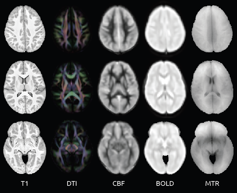
Sample slices from the multivariate atlas used as a basis for neuro-anatomical comparison


[Could not find the bibliography file(s)
Purpose: To evaluate the relationships between cerebral blood flow and other magnetic resonance (MR) imaging based measures such as fractional anisotropy, magnetic transfer ratio, cortical thickness and mean resting state BOLD signal in typically developing children.
Methods: Eighty-eight children aged 7-17 underwent pseudo-continuous arterial spin-labeled perfusion MRI (pCASL) [?] examinations along with anatomical (T1), diffusion tensor (DTI), magnetic transfer (MT) and BOLD resting state functional MRI (rs-fMRI) examinations. For each imaging modality, the ANTs [?] toolkit was used to create a modality-specific template from a subset (n=30) of the subjects. For non-scalar modalities, derived scalar images were used for template building. For pCASL the mean CBF image was used; for DTI the average diffusion weighted image was used; for rs-fMRI the mean BOLD image was used; and for MT the M0 image was used. Each modality-specific template was then registered to the T1 template to obtain a single multi-modality template (MMT). The T1 component of the MMT was then brain-masked, labeled, and three-tissue segmented using the Atropos segmentation tool [?]. For each subject, each modality was aligned to the corresponding component of the MMT for brain-masking and labeling. Intra-subject registrations were then performed to align all modalities to each subject’s T1 image. To provide a basis for comparison, a scalar metric was derived for each image modality. For pCASL the mean CBF was calculated; for T1 images, the cortical thickness was measured using the DiRECT method; fractional anisotropy was calculated from the DTI; the magnetization transfer ratio (MTR) was calculated from the MT images; and mean BOLD signal was calculated from the resting state fMRI data.
Results: Regularized canonical correlation analysis, as implemented in the sscan tool [?], was used to identify the relationship between CBF and each of the additional modalities. The analysis of each modality type is restricted to the most informative tissue type for that modality. For CBF, rs-fMRI and cortical thickness, only values in gray matter are examined, while only values in white matter are examined for FA and MTR.
Discussion: To the best of our knowledge, this is the first study to simultaneously compare CBF to cortical thickness, fractional anisotropy, magnetization transfer ratio and mean resting BOLD signal in a single population. In doing so, we hope to gain insight regarding the degree to which CBF provides statistically unique information in relation to these additional MR imaging modalities. Additionally, the development of the framework for analyzing these modalities provides a basis for future studies to explore the relationship between CBF and network based measures of both structural and functional connectivity.
Conclusion: The relationship between cortical thickness and Mean CBF (R2=0.4777) was the strongest of the metrics examined. In white matter, the MTR (R2=0.3126)  was stronger than FA (R2=0.1462). The mean BOLD (R2=0.1414) metric was the weakest.


Traumatic Brain Injury

This collaboration with the Moss Rehabilitation Research Institute at the Albert Einstein Healthcare Network involves the development of methologies for examining both structural and connective properties in the brains of TBI survivors.

Cardiac Medial Modeling

The aims of this collaborative project with the Computational Imaging Lab at the Pompeu Fabra University are to use a medial model of the myocardium to generate stronger shape priors for segmentation, richer features for shape analysis and a shape-based coordinate parameterization for the analysis of heart wall dynamics.

Structure-Specific fMRI Analysis

We are developing a new class of techniques that focus statistical analysis of functional neuroimaging data on specific structures such as the hippocampus, using the shape of the structures as the guide by which to combine information across subjects in ways that is more meaningful, sensitive and specific than in whole-brain normalization.

Multi-Atlas Segmentation

Objective

Segmentation, the problem of locating and outlining objects of interest in images, is a central problem in biomedical image analysis. It is the primary mechanism for quantifying the properties of anatomical structures and pathological formations using complex imaging data. With imaging used extensively across various fields of basic and clinical biomedical research, the value of accurate, reliable, and cost-effective segmentation cannot be understated. In brain research in particular, segmentation of MRI and other imaging modalities is crucial for studying the effects of behavior, disease, and treatment on brain anatomy and function. Despite years of research, automatic segmentation still generally underperforms manual segmentation in terms of reliability, and manual segmentation remains the gold standard in many problems. However, manual segmentation is often prohibitive, especially for large-scale studies or clinical trials. ‘The goal of our work is to develop, validate, and disseminate techniques that combine data from multiple sources in order to reduce the reliability gap between manual and automatic segmentation across a range of applications.