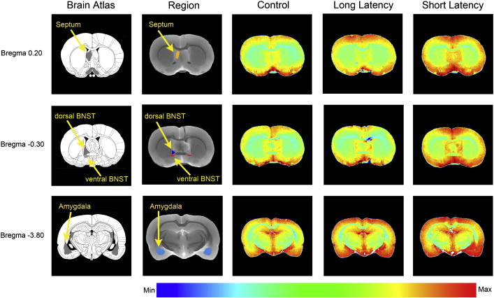
Swim stress produces relatively different magnitudes of Mn2 + enhancement in certain brain regions in rats with different coping strategies. The panels illustrate differences in Mn2 + enhancement in the septum, BNST, and amygdala in short latency (low stress coping) compared to long latency rats. Coronal sections (200 μm) from anterior to posterior are shown in the following order from left to right: Paxinos and Watson atlas section, the region(s) of interest analyzed, control rat, LL rat, and SL rat.
Abstract: Responses to acute stressors are determined in part by stress history. For example, a history of chronic stress results in facilitated responses to a novel stressor and this facilitation is considered to be adaptive. We previously demonstrated that repeated exposure of rats to the resident–intruder model of social stress results in the emergence of two subpopulations that are characterized by different coping responses to stress. The submissive subpopulation failed to show facilitation to a novel stressor and developed a passive strategy in the Porsolt forced swim test. Because a passive stress coping response has been implicated in the propensity to develop certain psychiatric disorders, understanding the unique circuitry engaged by exposure to a novel stressor in these subpopulations would advance our understanding of the etiology of stress-related pathology. An ex vivo functional imaging technique, manganese-enhanced magnetic resonance imaging (MEMRI), was used to identify and distinguish brain regions that are differentially activated by an acute swim stress (15 min) in rats with a history of social stress compared to controls. Specifically, Mn2 + was administered intracerebroventricularly prior to swim stress and brains were later imaged ex vivo to reveal activated structures. When compared to controls, all rats with a history of social stress showed greater activation in specific striatal, hippocampal, hypothalamic, and midbrain regions. The submissive subpopulation of rats was further distinguished by significantly greater activation in amygdala, bed nucleus of the stria terminalis, and septum, suggesting that these regions may form a circuit mediating responses to novel stress in individuals that adopt passive coping strategies. The finding that different circuits are engaged by a novel stressor in the two subpopulations of rats exposed to social stress implicates a role for these circuits in determining individual strategies for responding to stressors. Finally, these data underscore the utility of ex vivo MEMRI to identify and distinguish circuits engaged in behavioral responses.