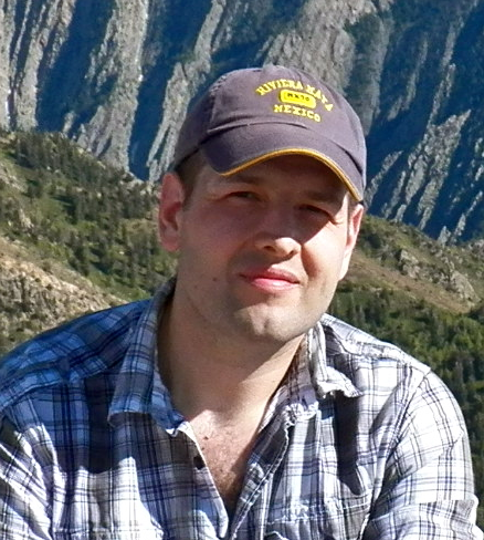
I am an Associate Professor in the Department of Radiology at the University of Pennsylvania and a member of the Bioengineering Graduate Group. My research focuses on developing novel computational methodologies for the analysis of biomedical imaging data. In my graduate work, performed under the direction of Stephen M. Pizer, Ph.D. at the University of North Carolina, I worked on the problems of finding shape representations suitable for statistical shape analysis with features derived from geometrical skeletons, and developed the continuous medial representation (cm-rep) approach. I have continued this research in my postgraduate work, developing new ways to use differential equations to solve the complex geometrical constraints that arise in skeleton-based shape analysis. I have been applying this methodology to the problems of shape analysis, shape-based normalization of brain structures, detailed structure-specific fMRI analysis, and tract-specific diffusion MRI analysis. As part of this structure-specific framework, I have developed a detailed atlas of the hippocampal region, derived from high-field, ultra high-resolution MRI images and dense histology stacks, along with techniques for leveraging this atlas in the analysis of in vivo MRI. My other work includes multi-atlas and shape-based segmentation algorithms, applied to problems in neuroimaging and cardiac imaging (including first-place finishes in segmentation grand challenges at MICCAI 2012 and MICCAI 2013), techniques for the analysis of functional and diffusion MRI data, groupwise registration, and many other image analysis topics. In addition to theoretical and applied image analysis research, I am deeply involved in efforts to make complex image analysis tools available broadly in the form of software applications. I supervise the development of ITK-SNAP, a mature multi-platform, open-source image segmentation software tool that is used widely in the field, and the companion tool Convert3D.
Publications
- Google Scholar
- PubMed
- In Press:
- P. A. Yushkevich, J. B. Pluta, H. Wang, L. Xie, S.-L. Ding, E. C. Gertje, L. Mancuso, D. Kliot, S. R. Das, and D. A. Wolk. Automated volumetry and regional thickness analysis of hippocampal subfields and medial temporal cortical structures in mild cognitive impairment. Human Brain Mapping, 2014. http://dx.doi.org/10.1002/hbm.22627
- L. Xie, J. Pluta, H. Wang, S. R. Das, L. Mancuso, D. Kliot, B. B. Avants, S.-L. Ding, D. A. Wolk, and P. A. Yushkevich. Automatic clustering and thickness measurement of anatomical variants of the human perirhinal cortex. In Medical Image Computing and Computer-Assisted Intervention – MICCAI 2014, pages 81–88. Springer, 2014. http://dx.doi.org/10.1007/978-3-319-10443-0_11
Grants
Software
- ITK-SNAP interactive medical image segmentation tool (collaboration with Guido Gerig, Ph.D. at the University of Utah)
- Convert3D command-line companion to ITK-SNAP
- Automatic Segmentation of Hippocampal Subfields (ASHS)
- CM-Rep tools for parametric deformable modeling of object skeletons
- PICSL Multi-Atlas Segmentation Tools (led by Hongzhi Wang, Ph.D.)
- HistoloZee histology and MRI co-registration tool (led by Daniel Adler)
- DTI-TK toolkit for diffusion MRI registration and tract-specific analysis (led by Hui Zhang, Ph.D., UCL)
