HistoloZee is a tool that integrates histology reconstruction, MRI co-registration, and manual segmentation tools in an easy-to-use and intuitive interface. HistoloZee permits real-time interaction with complex and large (multi-GB) histology datasets during the co-registration steps of histology reconstruction.
HistoloZee was developed by Daniel Adler under the supervision of Paul Yushkevich. Please contact us to request a pre-distribution copy of the software. HistoloZee is under active development and the release (v.03) currently run under Mac OS X (>10.6), with Linux and Windows versions forthcoming.
Basic reconstruction procedure for a stack of histology slides with accompanying reference imaging data (e.g. MRI):
- Slides from the tissue block are loaded and co-registered to each other to form an initial volumetric reconstruction. Sorting of slides and the setting of pixel size, slice thickness, and slice spacing are done during this stage.
- Reference MRI is loaded and aligned to the histology slide stack.
- Each slide is transformed to better match its corresponding cut-plane in the MRI.
… subsequent iteration of steps 2 & 3. Histology and MRI image transformations are combined harmoniously by a mapping all data into global, physical coordinates.
Main features:
- Formats: Loading of stacks of very large (>1GB) microscopy slides, including support for most common life-sciences imaging formats (implemented using OpenSlide) and loading of 3D reference image data (e.g. MRI) in NIfTI format
- Organization: Facility for sorting of slides and saving of reconstruction projects in XML format
- Visualization: Multi-scale viewing of slides and ability to load image overlays with transparency, thresholding, and live edge detection (OpenGL and GLSL shaders)
- 3D views: Linked cursors between orthogonal views of imaging data
- Manual affine slide registration: Tools for fine-scaled manual 2D-2D alignment of slides at any resolution
- Manual affine MRI registration: Tools for interactive 3D affine transformation of reference MRI to match histology slides
- Automated affine slide registration: GPU-accelerated metric computation (MSE, NCC, NMI) and transformations for alignment of slides to each other (2D-2D) and with reference MRI data (2D-3D)
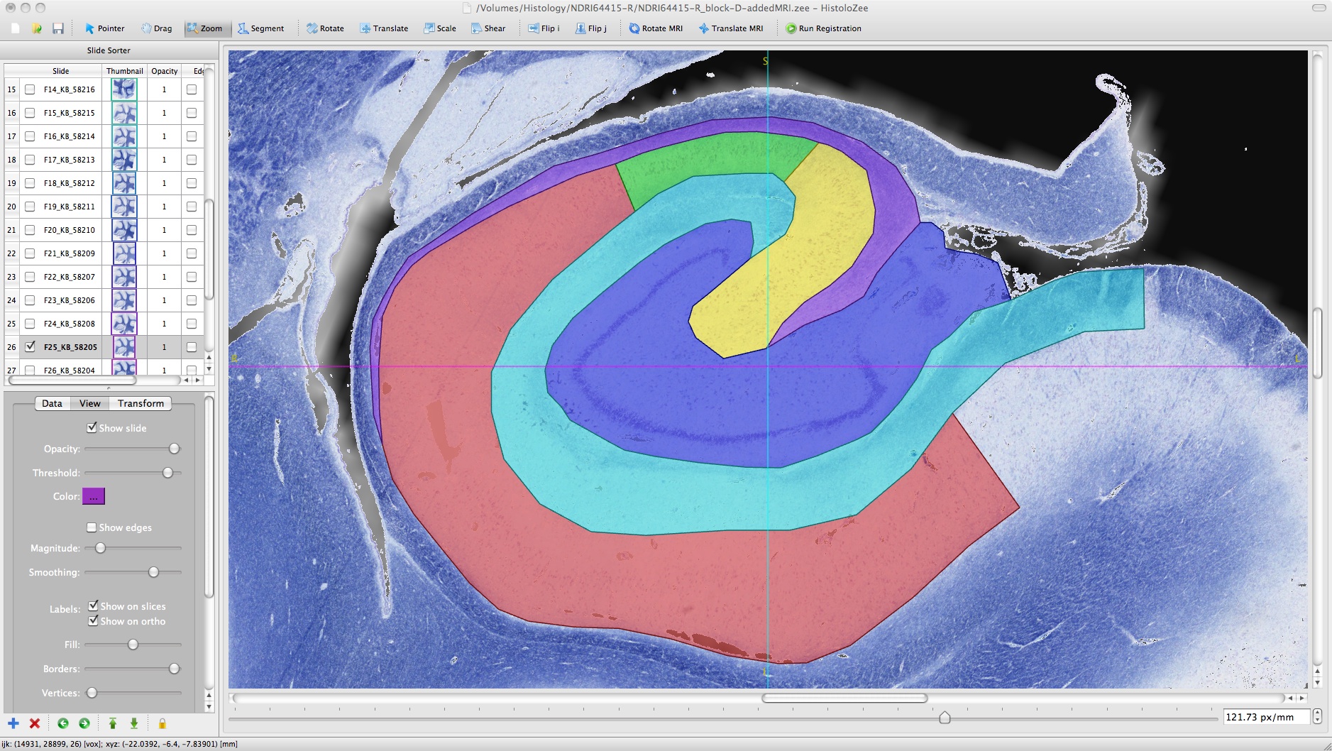
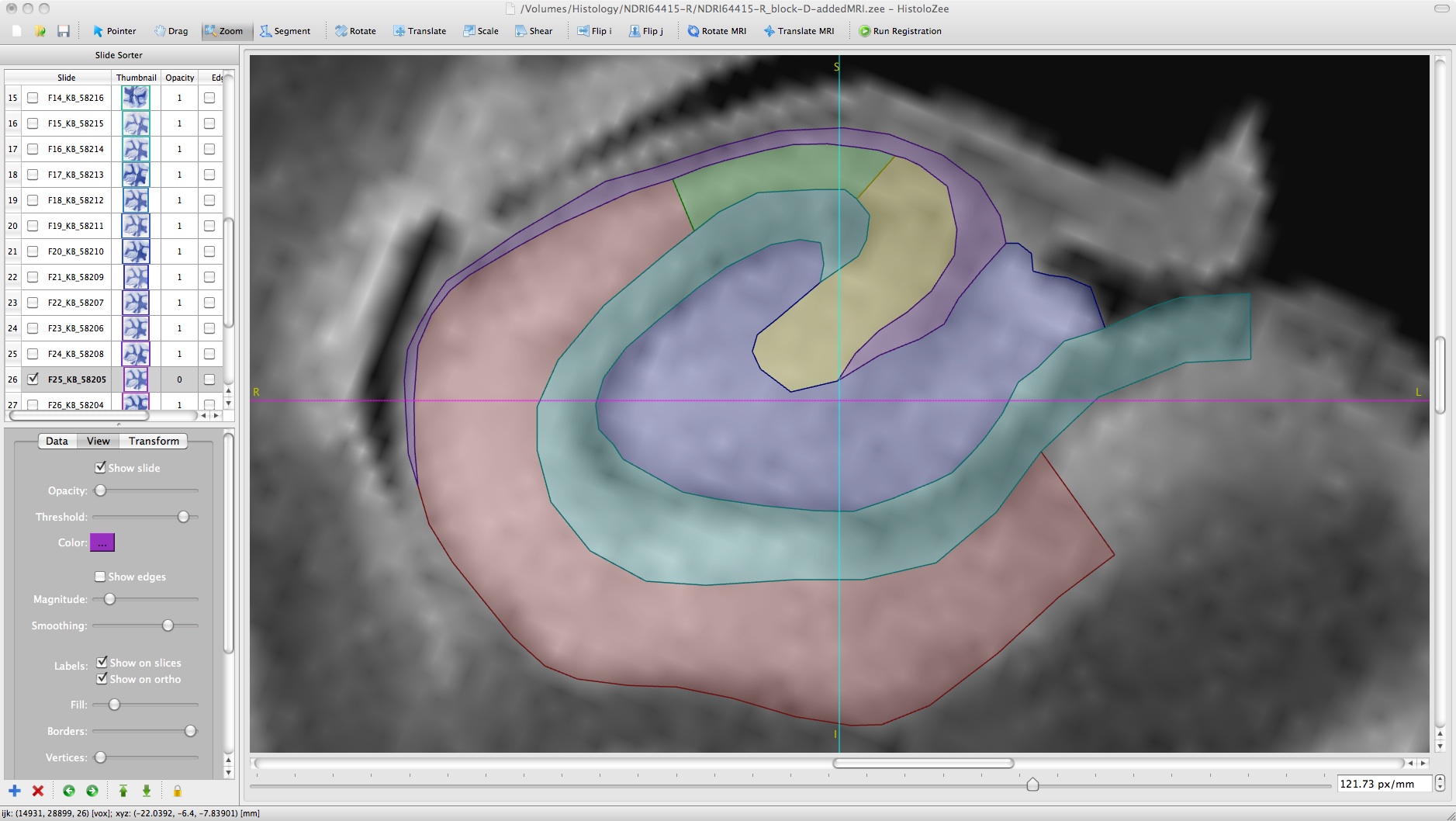
- Manual segmentation: Tools for delineation of regions using vector-graphics (“lossless”) label masks stored in SVG format
- Exporting: Reconstructed histology slide stacks can be exported to NIfTI format (or as an image series) at any resolution, while preserving the global coordinate mapping with MRI
Video tutorials:
Please visit our YouTube Playlist: HistoloZee Tutorials
Feel free to give us any feedback or suggestions on the tutorials and software!
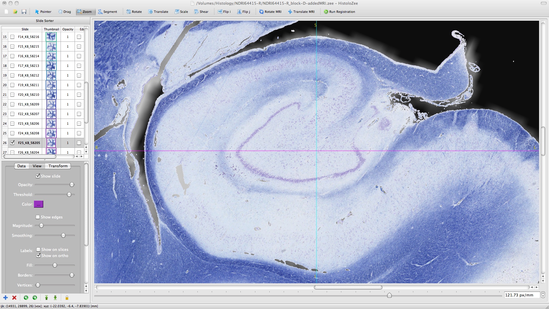
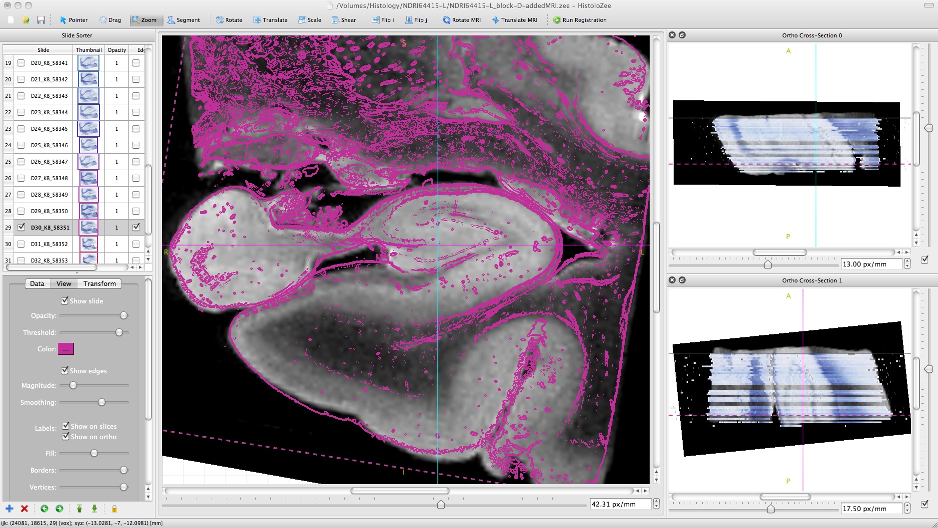
Screenshots:

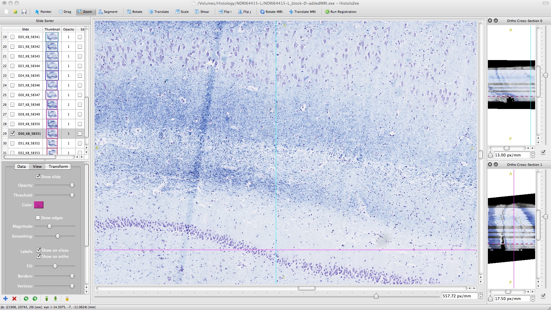
- Loading MRI reference image with reconstructed histology stack of human hippocampal formation.
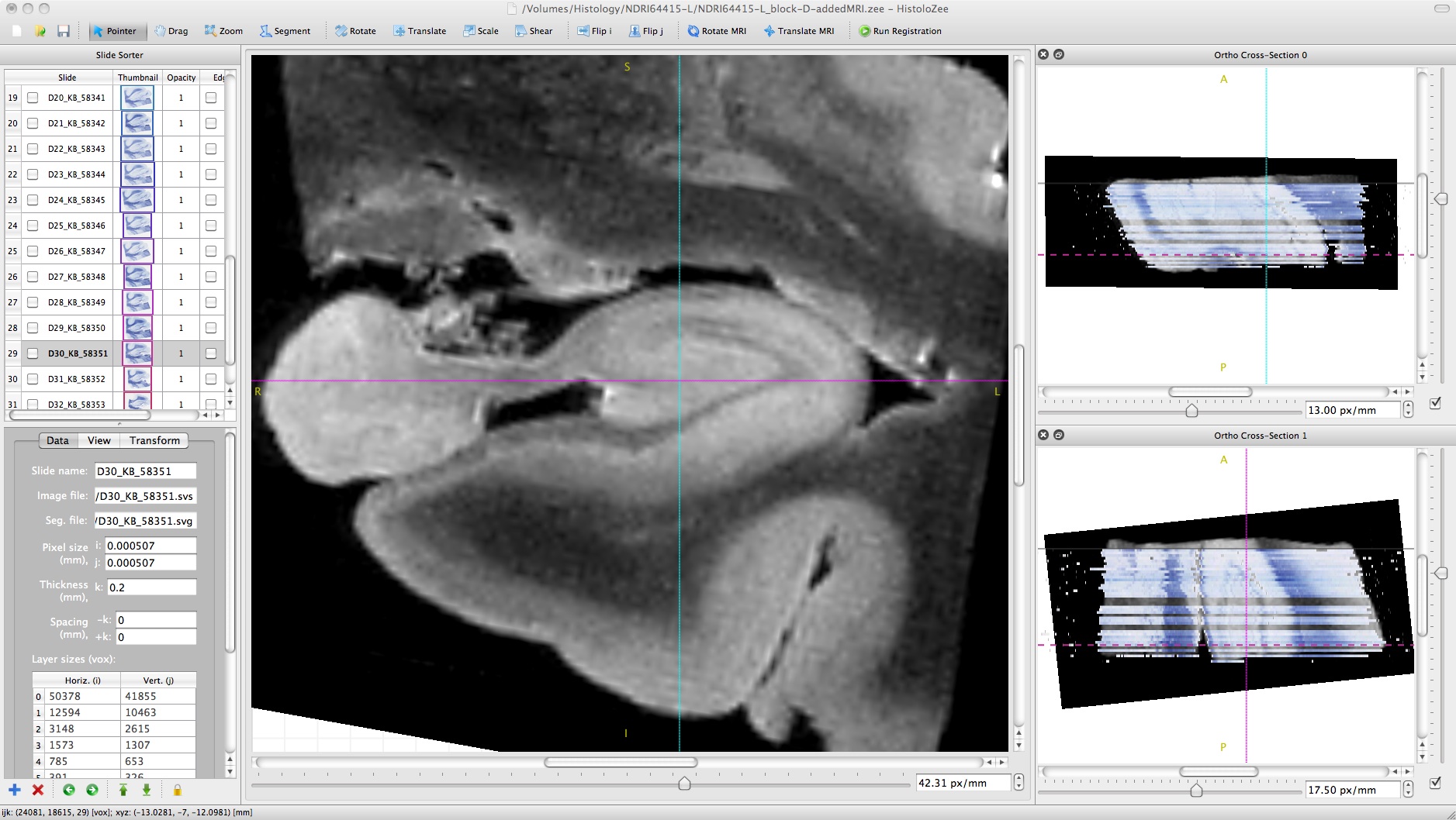

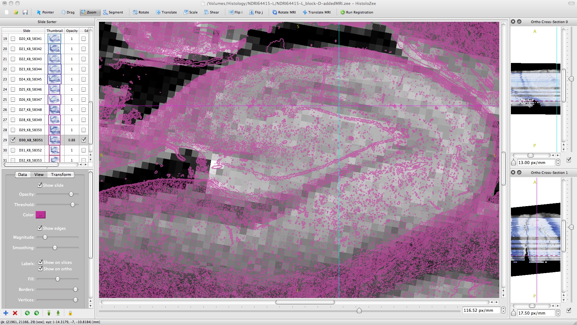
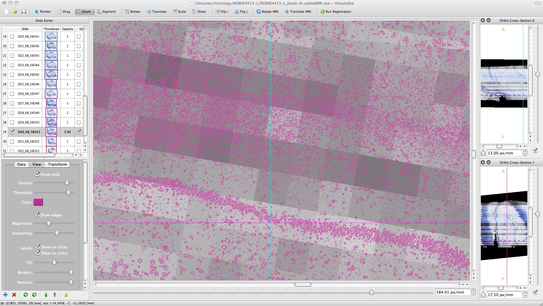
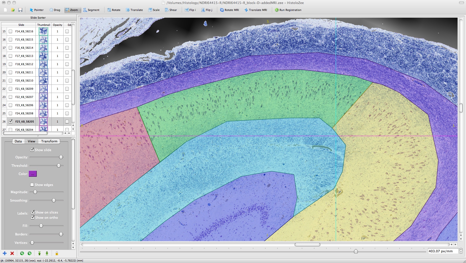
- Viewing histology slide with edge overlay atop MR reference image

- High resolution detail of cells