We are constructing a computational, probabilistic atlas of the human hippocampal formation (HF) from postmortem, high-resolution 9.4T MRI and densely acquired histology.
The human HF consists of several structures of the medial temporal lobe that play important roles in the declarative memory system. The HF is divided into cytoarchitecturally-defined subfields that serve different roles in the memory system and that are differentially affected by the processes of aging and neurological and psychiatric disorders.
MRI is the primary tool for biomarker discovery in the HF, since it is sensitive to small changes in shape/volume and robust to repeat measures over time. However, the HF substructures have complex anatomical structures that cannot be fully distinguished on routine in vivo MRI.
The atlas template image is being generated by normalizing postmortem MRI of multiple whole-HF tissue samples in a diffeomorphic registration framework. Histology, reconstructed in 3D from a subset of these samples, serves as the primary source for labeling structures in the atlas, including the subfields. When complete, our atlas will serve important roles in the hippocampal imaging field:
- Prior model for automatic in vivo MRI segmentation
- Guide development of manual MRI segmentation protocols
- Reference space for anatomical and functional imaging studies
- Anatomy learning resource
Image Acquisition Protocol
We acquire postmortem 3D MRI at 9.4T (~0.2mm isotropic) using a spin-echo multi-slice sequence from all HF tissue samples:
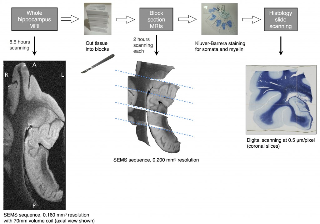
Postmortem MR and histological imaging protocol of the human hippocampal formation
A subset of the tissue samples is sectioned at 5μm thickness with 200μm spacing, stained using the Kluver-Barrera method (luxol fast blue + cresyl violet) for myelin and neuronal cell bodies, then digitally scanned at 0.5×0.5μm resolution.
Manual histology segmentation protocol
Subfields are manually segmented along the entire length of the HF in the histology slides. Boundaries between adjacent subfields are determined on the basis of cytoarchitectonic features (e.g. size, shape, and density of the neuronal somata), rather than using geometric or heuristic rules.
Histology slide and subfield label reconstructions
Histology images and labels are reconstructed in 3D and co-registered into the anatomical space of the matching postmortem MRI using a sequence of 2D and 3D affine and diffeomorphic registration steps:

Major steps of histology reconstruction pipeline shown schematically for one tissue block
For further details, please see our publication describing histology reconstruction in MRI space performed on one subject:
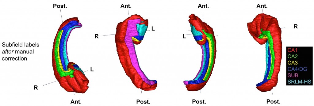
Hippocampal subfield labels derived from histology reconstructed in 3D after manual label refinement
This is the first work demonstrating hippocampal subfield labels in MRI space that are derived from ground-truth histological imaging. We are in the process of acquiring histology of more samples (normal and AD-affected cases) so that our atlas will capture variability of subfield distributions. We are also developing improved groupwise registration methodology for co-registering the MR images to create the atlas reference space.
We expect our postmortem atlas to serve as a valuable resource for future imaging studies by providing prior knowledge on the shapes and distributions of the subfields.