Tau-PET imaging and subregional atrophy in medial temporal lobe

We recently examined potential relationship between tau protein uptake in medial temporal lobe (MTL) and volumetric measures of MTL subregions in a small dataset of Alzheimer’s disease patients. Since tau protein accumulates in MTL in a characteristic spatiotempoal pattern as … Continue reading
Accounting for the Confound of Meninges in Segmenting Entorhinal and Perirhinal Cortices in T1-weighted MRI

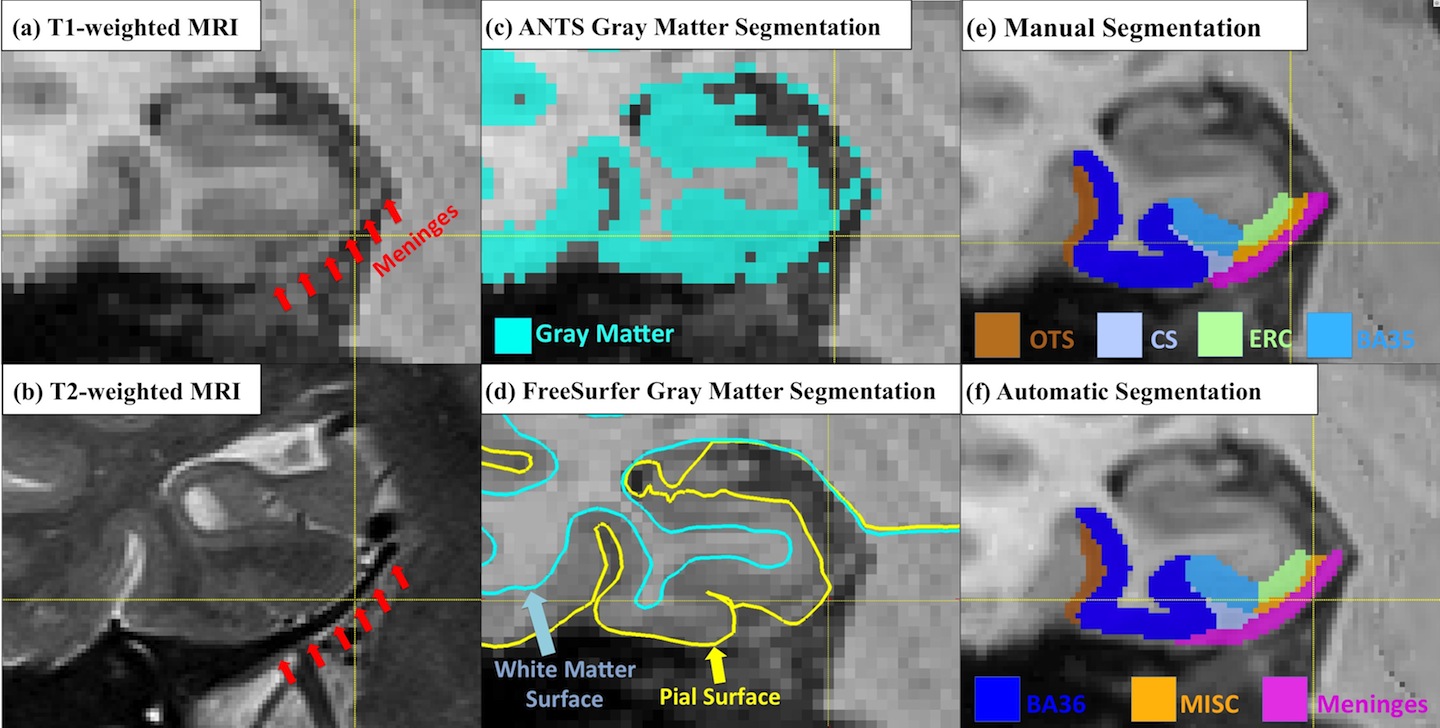
Quantification of medial temporal lobe (MTL) cortices, including entorhinal cortex (ERC) and perirhinal cortex (PRC), from in vivo MRI is desirable for studying the human memory system as well as in early diagnosis and monitoring of Alzheimer’s disease. However, ERC … Continue reading

Multi-Template Analysis of Human Perirhinal Cortex in Brain MRI: Explicitly Accounting for Anatomical Variability

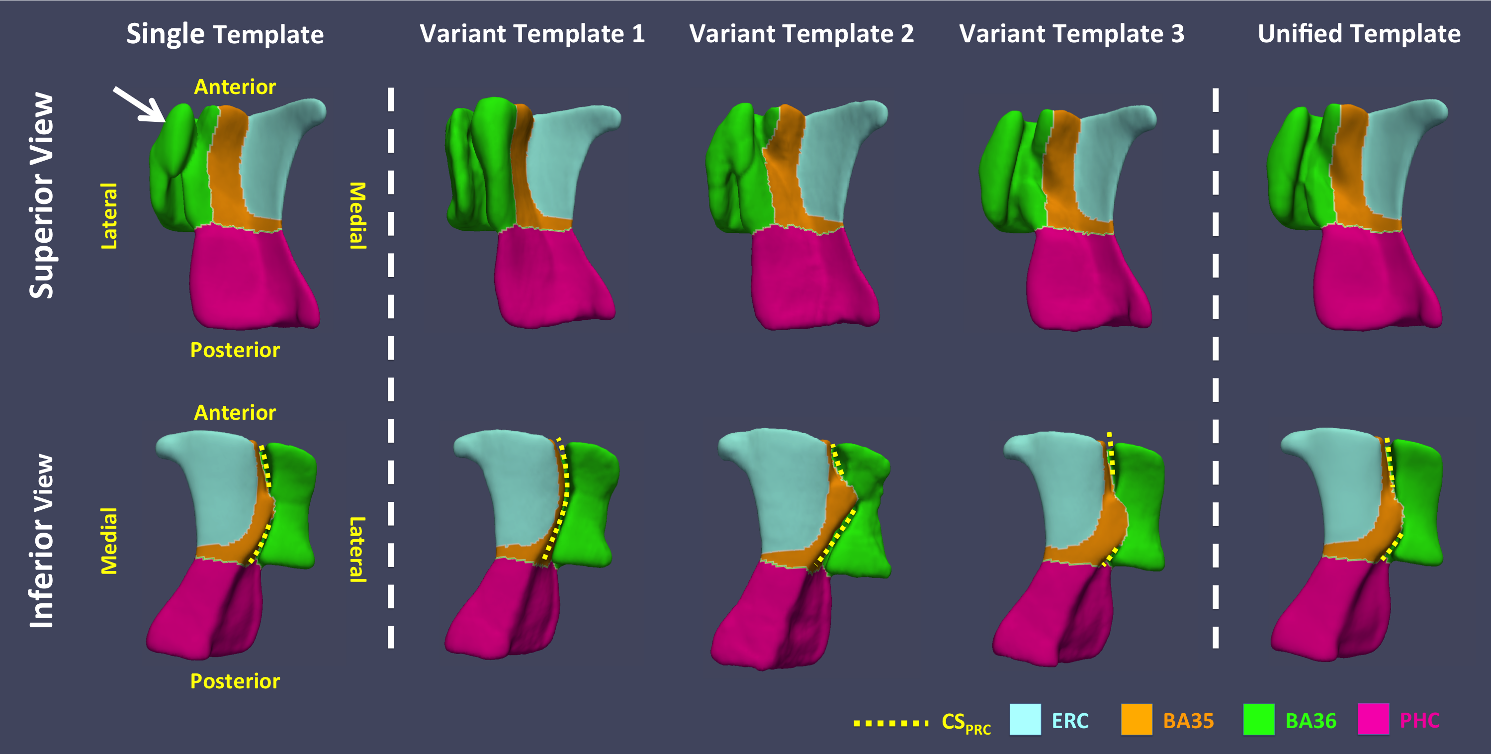
The human perirhinal cortex (PRC) plays critical roles in episodic and semantic memory and visual perception. The PRC consists of Brodmann areas 35 and 36 (BA35, BA36). In Alzheimer’s disease (AD), BA35 is the first cortical site affected by neurofibrillary … Continue reading

Medial temporal lobe subregional morphometry using high resolution MRI in Alzheimer’s Disease

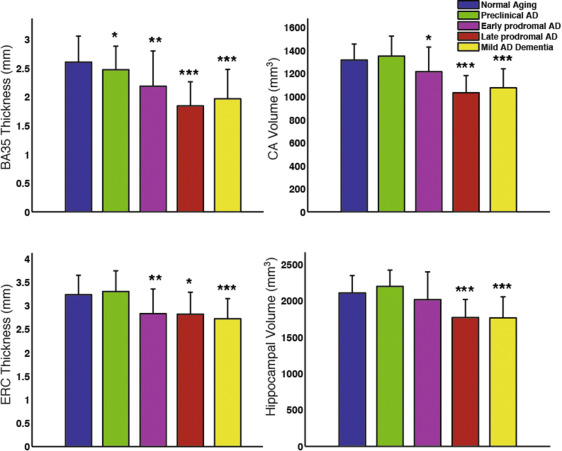
Recently published paper by Wolk et al. in Neurobiology of Aging examines the involvement of hippocampal subfields and medial temporal lobe (MTL) cortical subregions in different stages of Alzheimer’s disease (AD). The study used high-resolution T2-weighted MRI scans of the … Continue reading

Important milestone for the Penn ex vivo atlas of the human hippocampus formation

Daniel Adler’s MICCAI 2016 paper “Probabilistic atlas of the human hippocampus combining ex vivo MRI and histology” describes substantial progress on building a comprehensive atlas of the human hippocampus that combines ultra-high resolution ex vivo MRI and serial histological imaging. … Continue reading