The basis of mitral annuloplasty ring design has progressed from qualitative surgical intuition to experimental and theoretical analysis of annular geometry with quantitative imaging techniques. In this work, we present an automated 3D echocardiographic image analysis method that can be used to statistically assess variability in normal mitral annular geometry to support advancement in annuloplasty ring design.
3D patient-specific models of the mitral annulus are automatically generated from 3D echocardiographic images acquired from subjects with normal mitral valve structure and function. A mean 3D annular contour is computed, and principal component analysis is used to evaluate variability in normal annular shape.

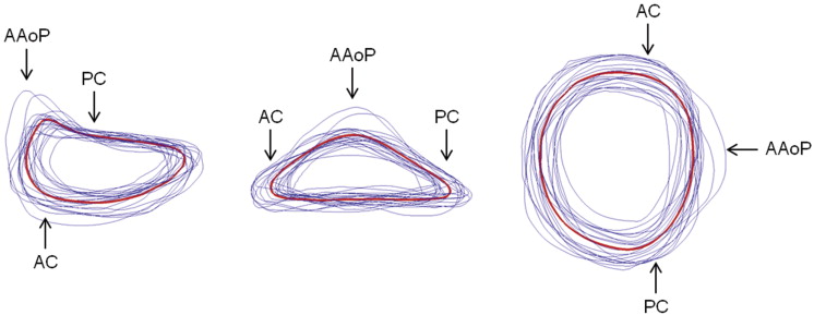
Mean (red) and individual (blue) 3D image-derived annular contours shown from three perspectives. (AAoP = anterior aortic peak; AC = anterior commissure; PC = posterior commissure.)

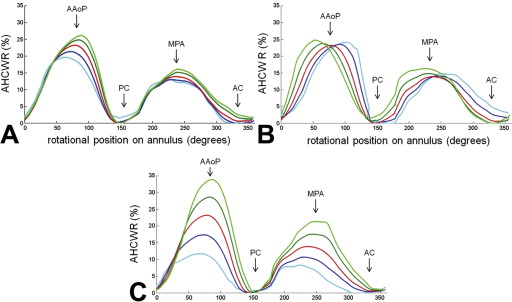
Annular height to intercommissural width ratio (AHCWR) as a function of rotational position on the annular contour. Each plot refers to one of three eigenmodes (A, mode 1; B, mode 2; C, mode 3) in shape variation obtained by principal component analysis. The red curve refers to the mean annular contour, the dark and light blue curves refer to +1 and +2 standard deviations from the mean, and the dark and light green curves refer to −1 and −2 standard deviations from the mean along a given eigenmode of shape variation. (AAoP = anterior aortic peak; AC = anterior commissure; MPA = midpoint of the posterior annulus; PC = posterior commissure.)
It is possible that a wider application of this analysis could provide information for a new generation of annuloplasty ring designs. All current designs are manufactured in a range of sizes with all sizes maintaining the same shape. With further study it may become apparent that to completely restore normal valve geometry, the next generation of saddle-shaped annuloplasty devices would best be created with subtle variations in shape as ring size increases.