Given the importance of image-based morphological assessment in the diagnosis and surgical treatment of aortic valve disease, there is considerable need to develop a standardized framework for 3D valve segmentation and shape representation. Towards this goal, this work integrates template-based medial modeling and multi-atlas label fusion techniques to automatically delineate and quantitatively describe aortic leaflet geometry in 3D echocardiographic images, a challenging task that has been explored only to a limited extent. The method makes use of expert knowledge of aortic leaflet image appearance, generates segmentations with consistent topology, and establishes a shape-based coordinate system on the aortic leaflets that enables standardized automated measurements.

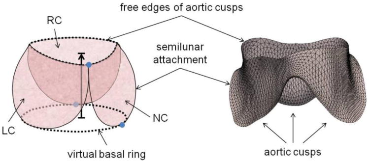
(Left) Schematic of the aortic cusps at systole. The valve orifice is the area enclosed by the cusp free edges, and the virtual basal ring connects the basal attachments of the cusps. Valve height is the distance between the virtual basal ring and valve orifice, shown by the black arrow pointing in the direction of blood flow. Three manually identified landmarks are shown in blue. (Right) The triangulated medial template of the aortic cusps used to initialize deformable modeling. (RC = right coronary cusp, LC = left coronary cusp, NC = noncoronary cusp)

A manual segmentation, consensus segmentation generated by label fusion, and fitted medial model of the aortic leaflets. The model-based segmentation is shown in red (right). The yellow arrow points in the direction of blood flow. (LC, RC, NC = left, right, and non-coronary cusps)
The development of this automated technique is a step towards creating a practical, informative tool for preoperative assessment of patient-specific aortic valve morphology.
Coming soon…
Segmentation of the complete aortic valve apparatus (cusps and root) with branching medial models.

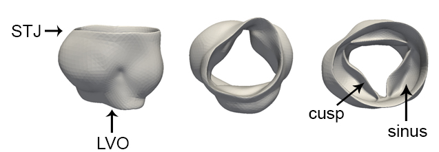
Aortic valve complex shown from three viewpoints: side (left), ventricular (center), and aortic (right) perspectives. (STJ = sinotubular junction, LVO = left ventricular outflow)