Here you can browse recent research posts. Mouseover the thumbnails to see a preview.
Tau-PET imaging and subregional atrophy in medial temporal lobe
Tau-PET imaging and subregional atrophy in medial temporal lobe
We recently examined potential relationship between tau protein uptake in medial temporal lobe (MTL) and volumetric measures of MTL subregions in a small dataset of Alzheimer’s disease patients. Since tau protein accumulates in MTL in a characteristic spatiotempoal pattern as … Continue reading
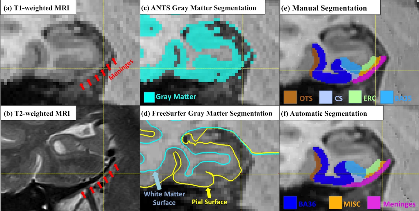
Accounting for the Confound of Meninges in Segmenting Entorhinal and Perirhinal Cortices in T1-weighted MRI
Accounting for the Confound of Meninges in Segmenting Entorhinal and Perirhinal Cortices in T1-weighted MRI
Quantification of medial temporal lobe (MTL) cortices, including entorhinal cortex (ERC) and perirhinal cortex (PRC), from in vivo MRI is desirable for studying the human memory system as well as in early diagnosis and monitoring of Alzheimer’s disease. However, ERC … Continue reading
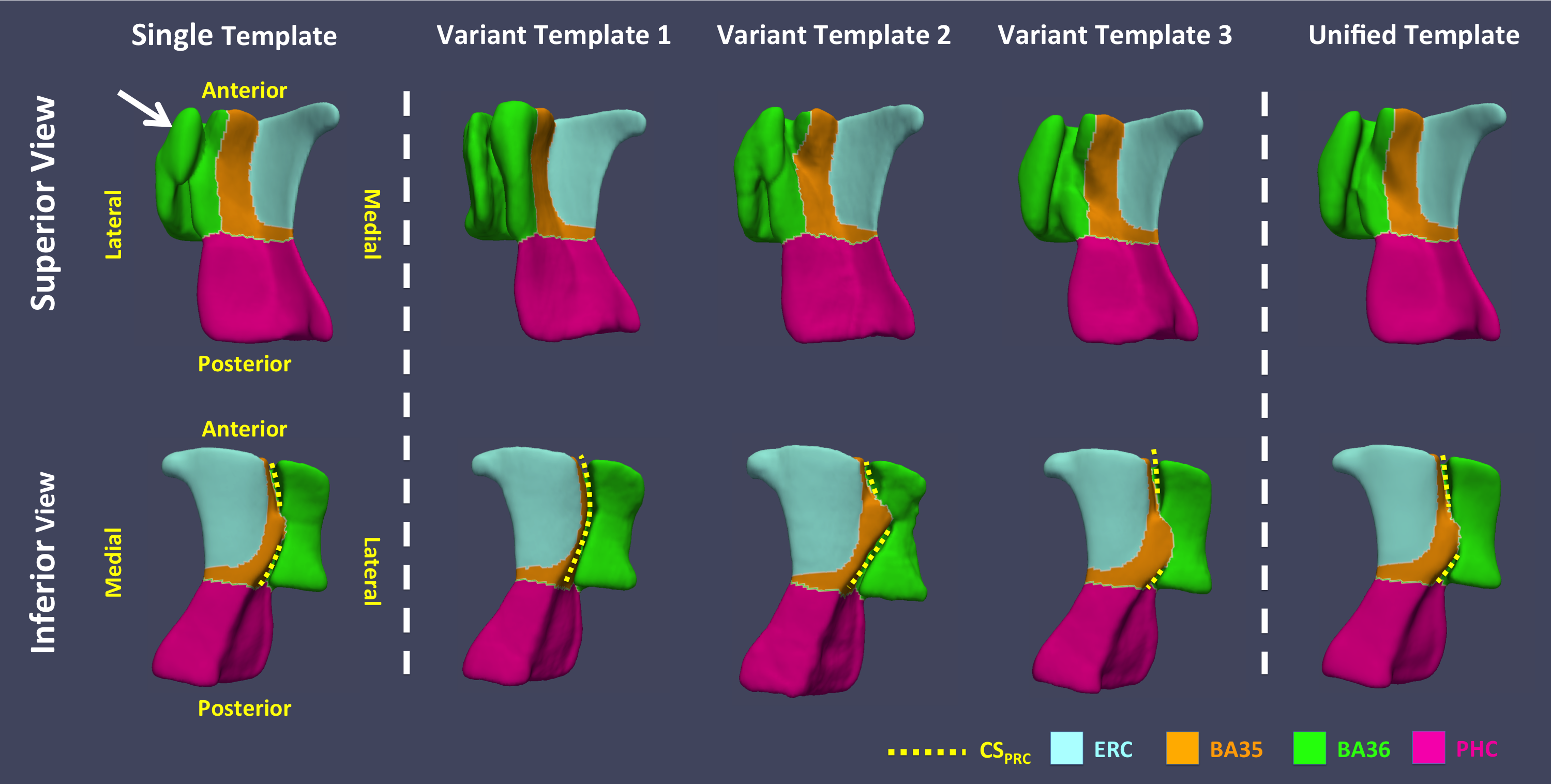
Multi-Template Analysis of Human Perirhinal Cortex in Brain MRI: Explicitly Accounting for Anatomical Variability
Multi-Template Analysis of Human Perirhinal Cortex in Brain MRI: Explicitly Accounting for Anatomical Variability
The human perirhinal cortex (PRC) plays critical roles in episodic and semantic memory and visual perception. The PRC consists of Brodmann areas 35 and 36 (BA35, BA36). In Alzheimer’s disease (AD), BA35 is the first cortical site affected by neurofibrillary … Continue reading
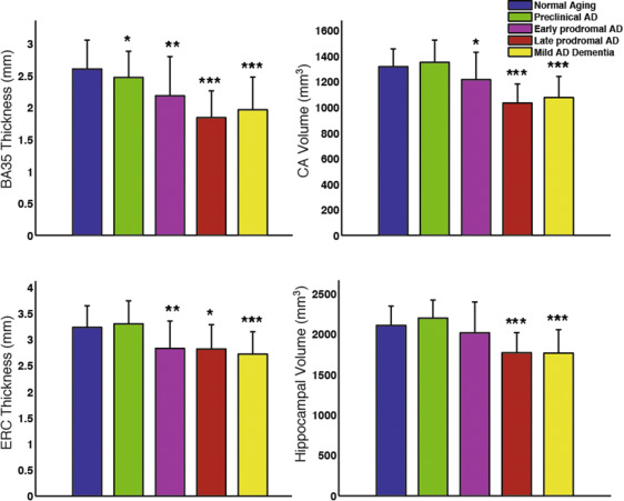
Medial temporal lobe subregional morphometry using high resolution MRI in Alzheimer’s Disease
Medial temporal lobe subregional morphometry using high resolution MRI in Alzheimer’s Disease
Recently published paper by Wolk et al. in Neurobiology of Aging examines the involvement of hippocampal subfields and medial temporal lobe (MTL) cortical subregions in different stages of Alzheimer’s disease (AD). The study used high-resolution T2-weighted MRI scans of the … Continue reading
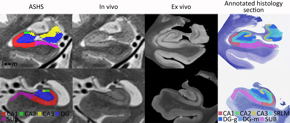
First comparison of in vivo/ ex vivo MRI scans of the human hippocampus
First comparison of in vivo/ ex vivo MRI scans of the human hippocampus
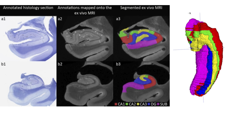
In our recent publication in Cerebral Cortex we compared in vivo and ex vivo MRI scans of the human hippocampus and evaluated to ability to match information between the two modalities. We were able to validate several of the features … Continue reading
Important milestone for the Penn ex vivo atlas of the human hippocampus formation
Important milestone for the Penn ex vivo atlas of the human hippocampus formation
Daniel Adler’s MICCAI 2016 paper “Probabilistic atlas of the human hippocampus combining ex vivo MRI and histology” describes substantial progress on building a comprehensive atlas of the human hippocampus that combines ultra-high resolution ex vivo MRI and serial histological imaging. … Continue reading
Relating brain anatomy and cognitive ability using a multivariate multimodal framework
Relating brain anatomy and cognitive ability using a multivariate multimodal framework
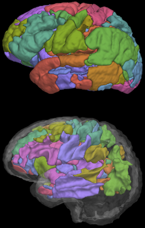
Linking structural neuroimaging data from multiple modalities to cognitive performance is an important challenge for cognitive neuroscience. In this study we examined the relationship between verbal fluency performance and neuroanatomy in 54 patients with frontotemporal degeneration (FTD) and 15 age-matched … Continue reading
Postmortem, probabilistic atlas of the human hippocampal formation
Postmortem, probabilistic atlas of the human hippocampal formation
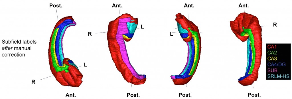
We are constructing a computational, probabilistic atlas of the human hippocampal formation (HF) from postmortem, high-resolution 9.4T MRI and densely acquired histology. The human HF consists of several structures of the medial temporal lobe that play important roles in the … Continue reading
ANTs Perfusion Processing
ANTs Perfusion Processing
We have recently updated ANTs to perform processing of perfusion ASL images. The pipeline is designed to be consistent with the antsCorticalThickness pipeline for computing cortical thickness and incorporates state-of-the-art methods for correcting for motion and other confounds. Learn more … Continue reading
Sparse Dimensionality Reduction for Medical Imaging
Sparse Dimensionality Reduction for Medical Imaging
The complex and high-dimensional nature of medical images makes them difficult to incorporate into traditional linear model-based statistical analysis. One way of simplifying analysis of medical images is to use a dimensionality reduction technique. We designed a dimensionality reduction technique … Continue reading
Sparse Regression for Medical Images
Sparse Regression for Medical Images
Our paper on sparse regression for predicting cognitive performance from cortical thickness was published at Information Processing in Medical Imaging (IPMI) 2013. We developed a method for performing regression on brain images in a way that respects the natural structure … Continue reading
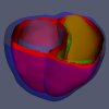
Segmentation of the aortic leaflets in 3D echocardiographic images
Segmentation of the aortic leaflets in 3D echocardiographic images
Given the importance of image-based morphological assessment in the diagnosis and surgical treatment of aortic valve disease, there is considerable need to develop a standardized framework for 3D valve segmentation and shape representation. Towards this goal, this work integrates template-based … Continue reading
Statistical analysis of normal mitral annular geometry
Statistical analysis of normal mitral annular geometry
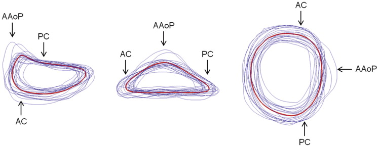
The basis of mitral annuloplasty ring design has progressed from qualitative surgical intuition to experimental and theoretical analysis of annular geometry with quantitative imaging techniques. In this work, we present an automated 3D echocardiographic image analysis method that can be … Continue reading
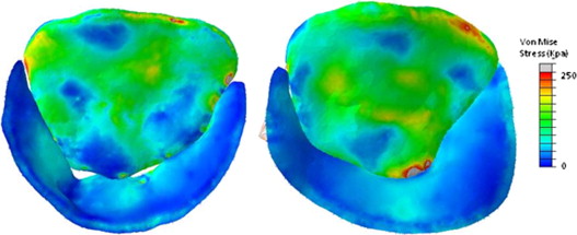
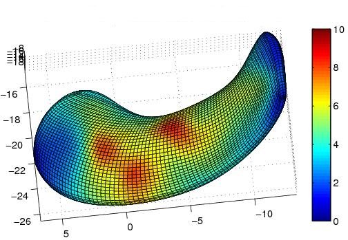
Prediction of stress distributions on image-derived models of the mitral leaflets
Prediction of stress distributions on image-derived models of the mitral leaflets
An integrated methodology for imaging, segmenting, modeling, and deriving computationally-predicted pressure-derived mitral leaflet stresses is presented and points the way towards intraoperative and periprocedural guidance from morphometric and stress modeling of the mitral valve. In vivo human mitral valves are … Continue reading
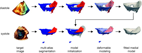
Fully automated segmentation of the mitral leaflets using multi-atlas label fusion and deformable medial modeling
Fully automated segmentation of the mitral leaflets using multi-atlas label fusion and deformable medial modeling
Comprehensive visual and quantitative analysis of in vivo human mitral valve morphology is central to the diagnosis and surgical treatment of mitral valve disease. Real-time 3D transesophageal echocardiography (3D TEE) is a practical, highly informative imaging modality for examining the … Continue reading
Semi-automated segmentation of the mitral leafelts in 3D echocardiographic images
Semi-automated segmentation of the mitral leafelts in 3D echocardiographic images
Precise 3D modeling of the mitral valve has the potential to improve our understanding of valve morphology, particularly in the setting of mitral regurgitation (MR). Toward this goal, we have developed a user-initialized algorithm for reconstructing valve geometry from transesophageal 3D echocardiographic (3DE) … Continue reading
7T fMRI R03 grant
7T fMRI R03 grant
We just found out that our R03 grant application for high-resolution BOLD fMRI at 7T has been recommended for funding. We have been anxiously waiting for the funding decision which was delayed by the government shutdown last year. Better late than … Continue reading
Functional connectivity within MTL
Functional connectivity within MTL
There is a ton of work looking at BOLD functional connectivity (FC) in Alzheimer’s Disease, and one consensus that has emerged is that this is a disconnection syndrome, with most studies finding weaker connectivity within the default mode network (DMN), … Continue reading
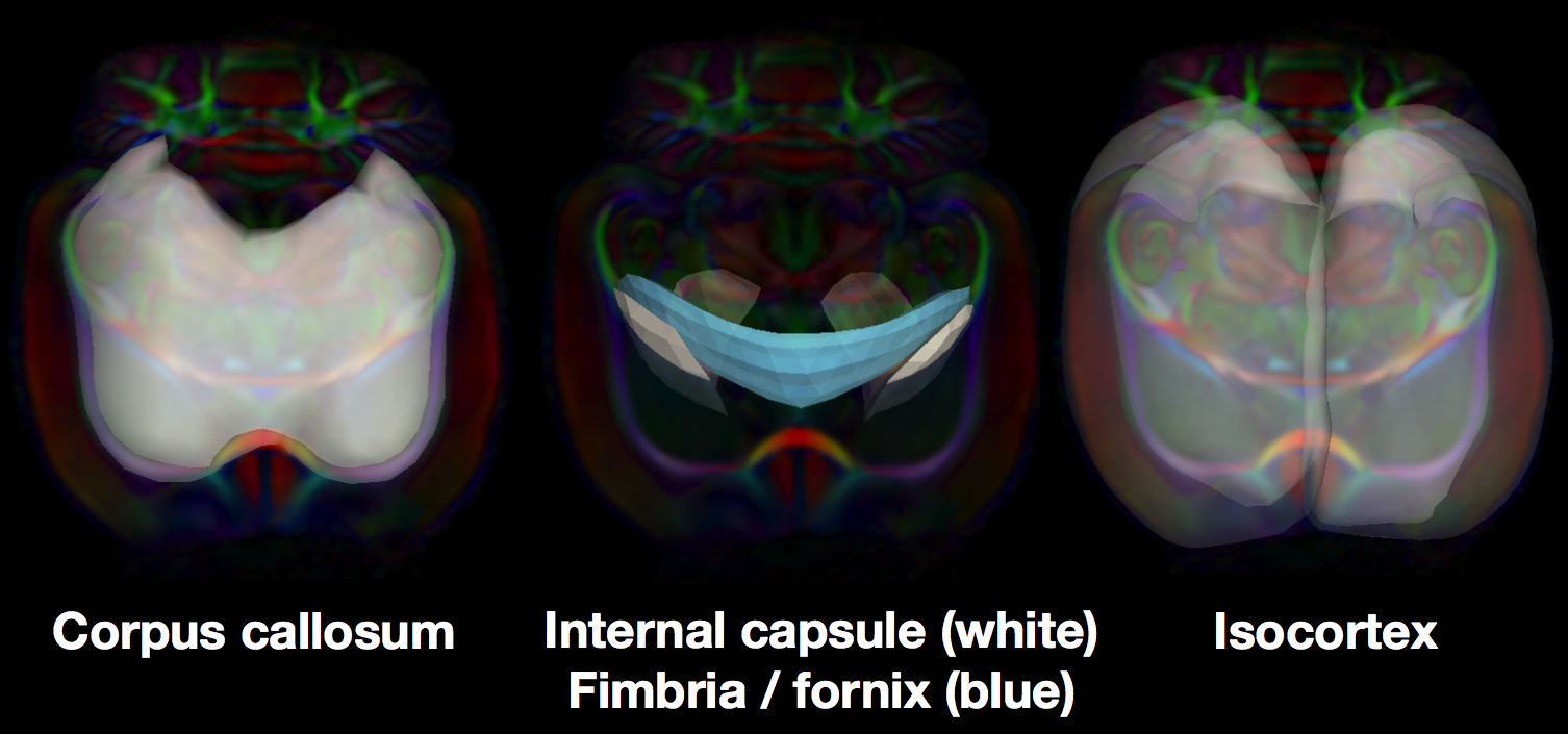
Structure Specific Analysis of white and gray matter in the rat brain
Structure Specific Analysis of white and gray matter in the rat brain
Tract specific Analysis (TSA) uses continuous medial modeling for structure- specific analysis of diffusion in sheet-like white matter tracts in the human brain. The medial geometry provides both a skeleton surface (the medial surface) and two “spoke” vectors from the … Continue reading
Tustison et al win the BRATS 2013 challenge
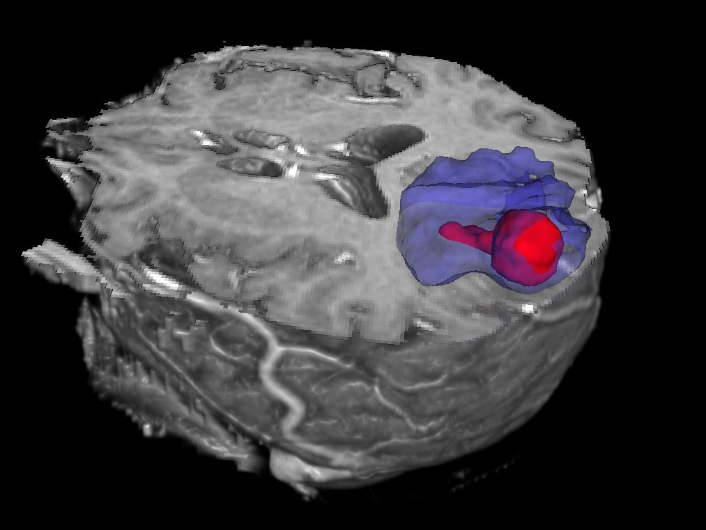
Tustison et al win the BRATS 2013 challenge
PICSL alumni Nick Tustison and colleagues Max Wintermark, Chris Durst, and Brian Avants finished in first place in the Multimodal Brain Tumor Segmentation challenge at the MICCAI conference (BRATS 2013): Given the success of random forest approaches for segmentation, particularly … Continue reading
Associations between children's socioeconomic status and prefrontal cortical thickness
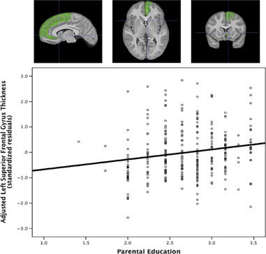
Associations between children's socioeconomic status and prefrontal cortical thickness
Childhood socioeconomic status (SES) predicts executive function performance and measures of prefrontal cortical function, but little is known about its anatomical correlates. Structural MRI and demographic data from a sample of 283 healthy children from the NIH MRI Study of … Continue reading
Manganese-enhanced magnetic resonance imaging (MEMRI) in rats with a history of repeated social stress.
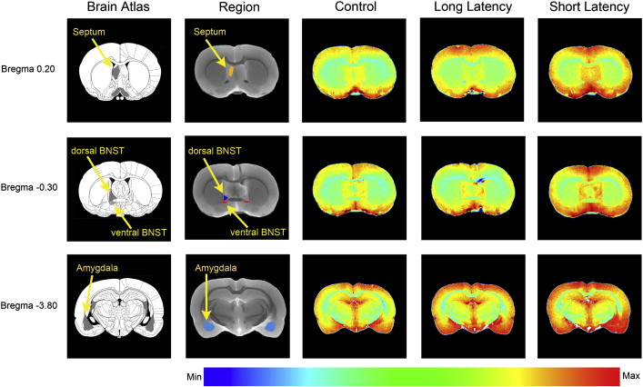
Manganese-enhanced magnetic resonance imaging (MEMRI) in rats with a history of repeated social stress.
Abstract: Responses to acute stressors are determined in part by stress history. For example, a history of chronic stress results in facilitated responses to a novel stressor and this facilitation is considered to be adaptive. We previously demonstrated that repeated … Continue reading
Relating Cerebral Blood Flow to Structural & Functional Metrics in Typically Developing Children
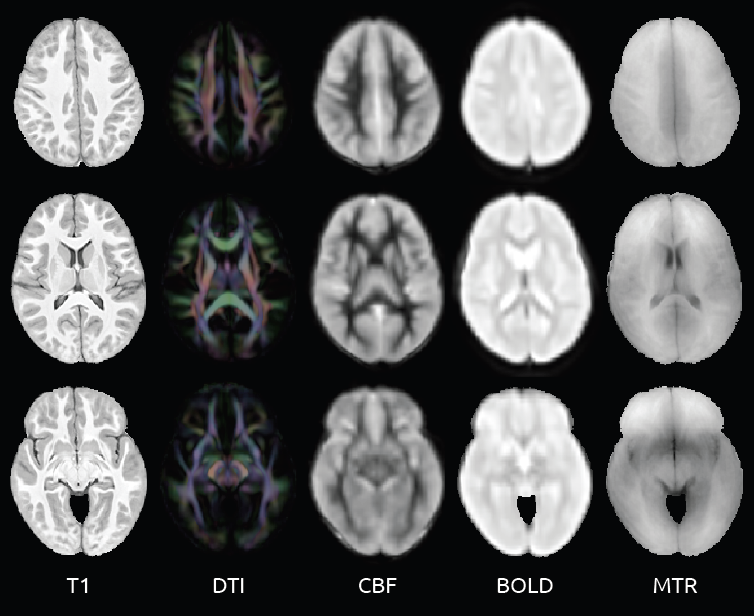
Relating Cerebral Blood Flow to Structural & Functional Metrics in Typically Developing Children
[Could not find the bibliography file(s) Purpose: To evaluate the relationships between cerebral blood flow and other magnetic resonance (MR) imaging based measures such as fractional anisotropy, magnetic transfer ratio, cortical thickness and mean resting state BOLD signal in typically … Continue reading

Traumatic Brain Injury
Traumatic Brain Injury
This collaboration with the Moss Rehabilitation Research Institute at the Albert Einstein Healthcare Network involves the development of methologies for examining both structural and connective properties in the brains of TBI survivors.
Cardiac Medial Modeling
Cardiac Medial Modeling
The aims of this collaborative project with the Computational Imaging Lab at the Pompeu Fabra University are to use a medial model of the myocardium to generate stronger shape priors for segmentation, richer features for shape analysis and a shape-based … Continue reading
Structure-Specific fMRI Analysis
Structure-Specific fMRI Analysis
We are developing a new class of techniques that focus statistical analysis of functional neuroimaging data on specific structures such as the hippocampus, using the shape of the structures as the guide by which to combine information across subjects in … Continue reading

Multi-Atlas Segmentation
Multi-Atlas Segmentation
Objective Segmentation, the problem of locating and outlining objects of interest in images, is a central problem in biomedical image analysis. It is the primary mechanism for quantifying the properties of anatomical structures and pathological formations using complex imaging data. … Continue reading