An integrated methodology for imaging, segmenting, modeling, and deriving computationally-predicted pressure-derived mitral leaflet stresses is presented and points the way towards intraoperative and periprocedural guidance from morphometric and stress modeling of the mitral valve.
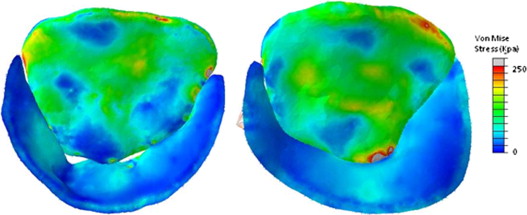
In vivo human mitral valves are imaged using real-time 3D transesophageal echocardiography, and volumetric images of the valve at mid systole are analyzed by user-initialized segmentation and 3D deformable modeling with continuous medial representation (cm-rep). The resulting models are loaded with physiologic pressures using finite element analysis. We present the regional leaflet stress distributions predicted in normal and diseased (regurgitant) mitral valves.
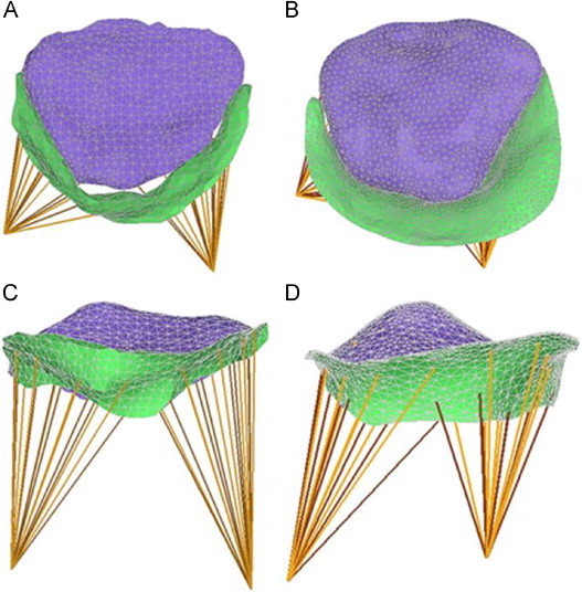
Finite element models of mid systolic diseased (A, C) and normal (B, D) mitral valves reconstructed from rt-3DTEE, in transvalvular (A, B) and oblique (C,D) views.
The ability to assess leaflet and chordal stresses in repaired valves will, with clinical experience, likely lead to improved surgical results by identifying patients with high stress valves in the early post-operative period. Such patients could either have re-repair or valve replacement before ever leaving the operating room, or could be subjected to closer post-operative clinical follow-up.
-
A. M. Pouch, C. Xu, P. A. Yushkevich, A. S. Jassar, M. Vergnat, J. H. Gorman 3rd, R. C. Gorman, C. M. Sehgal, and B. M. Jackson, “Semi-automated mitral valve morphometry and computational stress analysis using 3D ultrasound.,” J Biomech, vol. 45, iss. 5, p. 903–907, 2012.
[Bibtex]@ARTICLE{Pouch2012JB, author = {Pouch, Alison M. and Xu, Chun and Yushkevich, Paul A. and Jassar, Arminder S. and Vergnat, Mathieu and Gorman, 3rd, Joseph H and Gorman, Robert C. and Sehgal, Chandra M. and Jackson, Benjamin M.}, title = {{S}emi-automated mitral valve morphometry and computational stress analysis using 3{D} ultrasound.}, journal = {{J} {B}iomech}, year = {2012}, volume = {45}, pages = {903--907}, number = {5}, month = {Mar}, abstract = {In vivo human mitral valves (MV) were imaged using real-time 3D transesophageal echocardiography (rt-3DTEE), and volumetric images of the MV at mid-systole were analyzed by user-initialized segmentation and 3D deformable modeling with continuous medial representation, a compact representation of shape. The resulting MV models were loaded with physiologic pressures using finite element analysis (FEA). We present the regional leaflet stress distributions predicted in normal and diseased (regurgitant) MVs. Rt-3DTEE, semi-automated leaflet segmentation, 3D deformable modeling, and FEA modeling of the in vivo human MV is tenable and useful for evaluation of MV pathology.}, doi = {10.1016/j.jbiomech.2011.11.033}, institution = {{D}epartment of {B}ioengineering, {U}niversity of {P}ennsylvania, {P}hiladelphia, {PA}, {USA}.}, keywords = {Echocardiography, Three-Dimensional, methods; Echocardiography, Transesophageal, methods; Finite Element Analysis; Heart Valve Diseases, pathology/ultrasonography; Humans; Image Interpretation, Computer-Assisted, methods; Mitral Valve, pathology/ultrasonography; Models, Cardiovascular; Ultrasonics, methods}, language = {eng}, medline-pst = {ppublish}, owner = {alison}, pii = {S0021-9290(11)00715-9}, pmid = {22281408}, timestamp = {2014.02.27}, url = {http://dx.doi.org/10.1016/j.jbiomech.2011.11.033} }