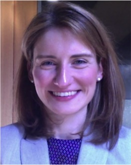
I am an Assistant Professor of Radiology and Bioengineering with interest in developing methods for computational image analysis, anatomical shape modeling, and interactive visualization to guide surgical interventions. Specifically, my work has focused on modeling heart valve morphology and dynamics in 3D echocardiography for risk stratification and planning of heart valve repair surgery. The goal is to use automated image analytics to predict surgical outcomes, to inform development of surgical guidelines, and to advance minimally invasive therapies for the treatment of cardiovascular disease.
Research Areas
3D/4D segmentation and modeling of heart valves in echocardiographic images with applications to surgical treatment of valvular regurgitation
- Semi-automated and fully automated segmentation of the mitral leaflets in 3D echocardiographic images
- Segmentation of the aortic valve apparatus, including the aortic root and tricuspid and bicuspid aortic valves
Integration of methods for image segmentation, shape representation, and biomechanical modeling
- Prediction of mitral leaflet stress distributions using patient-specific image-derived models of the mitral valve
Meshing and analysis of 3D anatomical shapes
- Statistical analysis of “normal” mitral annular geometry
- Automated meshing of the placenta in 3D ultrasound
3D printing of anatomical shapes in medical images
Education
University of Pennsylvania, 2018
- Certificate in Biomedical Informatics
University of Pennsylvania, 2007 – 2013
- Ph.D. in Bioengineering
- HHMI-NIBIB Interfaces Program in Biomedical Imaging and Informational Sciences
The College of William and Mary, 2003 – 2007
- B.S. in Physics with a dual concentration in Anthropology
Links
Research Gate | PubMed | Google Scholar
Peer-reviewed journal and conference publications
-
A. S. Jassar, M. M. Levack, R. Solorzano, A. M. Pouch, G. Ferrari, A. Cheung, V. A. Ferrari, J. H. Gorman 3rd, R. Gorman, and B. M. Jackson, “Feasibility of In Vivo Human Aortic Valve Modeling Using Real-Time Three-Dimensional Echocardiography.,” Ann Thorac Surg, 2014.
[Bibtex]@ARTICLE{Jassar2014ATS, author = {Jassar, Arminder S. and Levack, Melissa M. and Solorzano, Ricardo D. and Pouch, Alison M. and Ferrari, Giovanni and Cheung, Albert T. and Ferrari, Victor A. and Gorman, 3rd, Joseph H and Gorman, Robert C. and Jackson, Benjamin M.}, title = {{F}easibility of {I}n {V}ivo {H}uman {A}ortic {V}alve {M}odeling {U}sing {R}eal-{T}ime {T}hree-{D}imensional {E}chocardiography.}, journal = {{A}nn {T}horac {S}urg}, year = {2014}, month = {Feb}, abstract = {Surgical techniques for aortic valve (AV) repair are directed toward restoring normal structural relationships in the aortic root and rely on detailed assessment of root and valve anatomy. Noninvasive three-dimensional (3D) imaging and modeling may assist in patient selection and operative planning.Transesophageal real-time 3D echocardiographic images of 5 patients with normal AV were acquired. The aortic root and the annulus were manually segmented at end diastole using a 36-point rotational template. The AV leaflets and the coaptation zone were manually segmented in parallel 1-mm cross sections. Quantitative 3D models of the AV and root were generated and used to measure standard anatomic parameters and were compared to conventional two-dimensional echocardiographic measurements. All measurements are given as mean ± SD.Annular, sinus, and sinotubular junction areas were 4.1 ± 0.6 cm(2), 7.5 ± 1.2 cm(2), and 3.9 ± 1.0 cm(2), respectively. Root diameters (measured in three locations) by 3D model inspection and two-dimensional echocardiography measurement correlated (R(2) = 0.75). Noncoapted areas of the left, right, and noncoronary leaflets were 1.9 ± 0.2 cm(2), 1.6 ± 0.3 cm(2), and 1.6 ± 0.3 cm(2), respectively. Mean coaptation areas for the left-right, left-noncoronary, and right-noncoronary coaptation zones were 87.7 ± 36.9 mm(2), 69.9 ± 20.7 mm(2), and 114.2 ± 23.0 mm(2), respectively. The mean ratio of noncoapted leaflet area to annular area was 1.3 ± 0.2.High-resolution 3D models of the in vivo normal human aortic root and valve were generated using 3D echocardiography. Quantitative 3D models and analysis may assist in characterization of pathology and decision making for AV repair.}, doi = {10.1016/j.athoracsur.2013.12.017}, institution = {{D}epartment of {S}urgery, {U}niversity of {P}ennsylvania, {P}hiladelphia, {P}ennsylvania. {E}lectronic address: benjamin.jackson@uphs.upenn.edu.}, language = {eng}, medline-pst = {aheadofprint}, owner = {alison}, pii = {S0003-4975(13)02852-X}, pmid = {24518577}, timestamp = {2014.02.27}, url = {http://dx.doi.org/10.1016/j.athoracsur.2013.12.017} }
-
E. K. Shang, E. Lai, A. M. Pouch, R. Hinmon, R. C. Gorman, J. H. Gorman 3rd, C. Sehgal, G. Ferrari, J. E. Bavaria, and B. Jackson, “Validation of semiautomated and locally resolved aortic wall thickness measurements from computed tomography.,” J Vasc Surg, 2014.
[Bibtex]@ARTICLE{Shang2014JVS, author = {Shang, Eric K. and Lai, Eric and Pouch, Alison M. and Hinmon, Robin and Gorman, Robert C. and Gorman, 3rd, Joseph H and Sehgal, Chandra M. and Ferrari, Giovanni and Bavaria, Joseph E. and Jackson, Benjamin M.}, title = {{V}alidation of semiautomated and locally resolved aortic wall thickness measurements from computed tomography.}, journal = {{J} {V}asc {S}urg}, year = {2014}, month = {Jan}, abstract = {Aortic wall thickness (AWT) is important for anatomic description and biomechanical modeling of aneurysmal disease. However, no validated, noninvasive method for measuring AWT exists. We hypothesized that semiautomated image segmentation algorithms applied to computed tomography angiography (CTA) can accurately measure AWT.Aortic samples from 10 patients undergoing open thoracoabdominal aneurysm repair were taken from sites of the proximal or distal anastomosis, or both, yielding 13 samples. Aortic specimens were fixed in formalin, embedded in paraffin, and sectioned. After staining with hematoxylin and eosin and Masson's trichrome, sections were digitally scanned and measured. Patients' preoperative CTA Digital Imaging and Communications in Medicine (DICOM; National Electrical Manufacturers Association, Rosslyn, Va) images were segmented into luminal, inner arterial, and outer arterial surfaces with custom algorithms using active contours, isoline contour detection, and texture analysis. AWT values derived from image data were compared with measurements of corresponding pathologic specimens.AWT determined by CTA averaged 2.33 ± 0.66 mm (range, 1.52-3.55 mm), and the AWT of pathologic specimens averaged 2.36 ± 0.75 mm (range, 1.51-4.16 mm). The percentage difference between pathologic specimens and CTA-determined AWT was 9.5\% ± 4.1\% (range, 1.8\%-16.7\%). The correlation between image-based measurements and pathologic measurements was high (R = 0.935). The 95\% limits of agreement computed by Bland-Altman analysis fell within the range of -0.42 and 0.42 mm.Semiautomated analysis of CTA images can be used to accurately measure regional and patient-specific AWT, as validated using pathologic ex vivo human aortic specimens. Descriptions and reconstructions of aortic aneurysms that incorporate locally resolved wall thickness are feasible and may improve future attempts at biomechanical analyses.}, doi = {10.1016/j.jvs.2013.11.065}, institution = {{D}epartment of {S}urgery, {U}niversity of {P}ennsylvania, {P}hiladelphia, {P}a; {D}ivision of {V}ascular {S}urgery and {E}ndovascular {T}herapy, {U}niversity of {P}ennsylvania, {P}hiladelphia, {P}a. {E}lectronic address: benjamin.jackson@uphs.upenn.edu.}, language = {eng}, medline-pst = {aheadofprint}, owner = {alison}, pii = {S0741-5214(13)02160-5}, pmid = {24388698}, timestamp = {2014.02.27}, url = {http://dx.doi.org/10.1016/j.jvs.2013.11.065} }
-
A. M. Pouch, M. Vergnat, J. R. McGarvey, G. Ferrari, B. M. Jackson, C. M. Sehgal, P. A. Yushkevich, R. C. Gorman, and J. Gorman 3rd, “Statistical assessment of normal mitral annular geometry using automated three-dimensional echocardiographic analysis.,” Ann Thorac Surg, vol. 97, iss. 1, p. 71–77, 2014.
[Bibtex]@ARTICLE{Pouch2014ATS, author = {Pouch, Alison M. and Vergnat, Mathieu and McGarvey, Jeremy R. and Ferrari, Giovanni and Jackson, Benjamin M. and Sehgal, Chandra M. and Yushkevich, Paul A. and Gorman, Robert C. and Gorman, 3rd, Joseph H}, title = {{S}tatistical assessment of normal mitral annular geometry using automated three-dimensional echocardiographic analysis.}, journal = {{A}nn {T}horac {S}urg}, year = {2014}, volume = {97}, pages = {71--77}, number = {1}, month = {Jan}, abstract = {The basis of mitral annuloplasty ring design has progressed from qualitative surgical intuition to experimental and theoretical analysis of annular geometry with quantitative imaging techniques. In this work, we present an automated three-dimensional (3D) echocardiographic image analysis method that can be used to statistically assess variability in normal mitral annular geometry to support advancement in annuloplasty ring design.Three-dimensional patient-specific models of the mitral annulus were automatically generated from 3D echocardiographic images acquired from subjects with normal mitral valve structure and function. Geometric annular measurements including annular circumference, annular height, septolateral diameter, intercommissural width, and the annular height to intercommissural width ratio were automatically calculated. A mean 3D annular contour was computed, and principal component analysis was used to evaluate variability in normal annular shape.The following mean ± standard deviations were obtained from 3D echocardiographic image analysis: annular circumference, 107.0 ± 14.6 mm; annular height, 7.6 ± 2.8 mm; septolateral diameter, 28.5 ± 3.7 mm; intercommissural width, 33.0 ± 5.3 mm; and annular height to intercommissural width ratio, 22.7\% ± 6.9\%. Principal component analysis indicated that shape variability was primarily related to overall annular size, with more subtle variation in the skewness and height of the anterior annular peak, independent of annular diameter.Patient-specific 3D echocardiographic-based modeling of the human mitral valve enables statistical analysis of physiologically normal mitral annular geometry. The tool can potentially lead to the development of a new generation of annuloplasty rings that restore the diseased mitral valve annulus back to a truly normal geometry.}, doi = {10.1016/j.athoracsur.2013.07.096}, institution = {{G}orman {C}ardiovascular {R}esearch {G}roup, {U}niversity of {P}ennsylvania, {P}hiladelphia, {P}ennsylvania; {D}epartment of {S}urgery, {U}niversity of {P}ennsylvania, {P}hiladelphia, {P}ennsylvania. {E}lectronic address: gormanj@uphs.upenn.edu.}, language = {eng}, medline-pst = {ppublish}, owner = {alison}, pii = {S0003-4975(13)01709-8}, pmid = {24090576}, timestamp = {2014.02.27}, url = {http://dx.doi.org/10.1016/j.athoracsur.2013.07.096} }
-
A. M. Pouch, H. Wang, M. Takabe, B. M. Jackson, J. Gorman 3rd, R. C. Gorman, P. A. Yushkevich, and C. M. Sehgal, “Fully automatic segmentation of the mitral leaflets in 3D transesophageal echocardiographic images using multi-atlas joint label fusion and deformable medial modeling.,” Med Image Anal, vol. 18, iss. 1, p. 118–129, 2014.
[Bibtex]@ARTICLE{Pouch2014MIA, author = {Pouch, A. M. and Wang, H. and Takabe, M. and Jackson, B. M. and Gorman, 3rd, JH and Gorman, R. C. and Yushkevich, P. A. and Sehgal, C. M.}, title = {{F}ully automatic segmentation of the mitral leaflets in 3{D} transesophageal echocardiographic images using multi-atlas joint label fusion and deformable medial modeling.}, journal = {{M}ed {I}mage {A}nal}, year = {2014}, volume = {18}, pages = {118--129}, number = {1}, month = {Jan}, abstract = {Comprehensive visual and quantitative analysis of in vivo human mitral valve morphology is central to the diagnosis and surgical treatment of mitral valve disease. Real-time 3D transesophageal echocardiography (3D TEE) is a practical, highly informative imaging modality for examining the mitral valve in a clinical setting. To facilitate visual and quantitative 3D TEE image analysis, we describe a fully automated method for segmenting the mitral leaflets in 3D TEE image data. The algorithm integrates complementary probabilistic segmentation and shape modeling techniques (multi-atlas joint label fusion and deformable modeling with continuous medial representation) to automatically generate 3D geometric models of the mitral leaflets from 3D TEE image data. These models are unique in that they establish a shape-based coordinate system on the valves of different subjects and represent the leaflets volumetrically, as structures with locally varying thickness. In this work, expert image analysis is the gold standard for evaluating automatic segmentation. Without any user interaction, we demonstrate that the automatic segmentation method accurately captures patient-specific leaflet geometry at both systole and diastole in 3D TEE data acquired from a mixed population of subjects with normal valve morphology and mitral valve disease.}, doi = {10.1016/j.media.2013.10.001}, institution = {{D}epartment of {B}ioengineering, {U}niversity of {P}ennsylvania, {P}hiladelphia, {PA}, {U}nited {S}tates; {G}orman {C}ardiovascular {R}esearch {G}roup, {U}niversity of {P}ennsylvania, {P}hiladelphia, {PA}, {U}nited {S}tates. {E}lectronic address: pouch@seas.upenn.edu.}, language = {eng}, medline-pst = {ppublish}, owner = {alison}, pii = {S1361-8415(13)00142-4}, pmid = {24184435}, timestamp = {2014.02.27}, url = {http://dx.doi.org/10.1016/j.media.2013.10.001} }
- A. M. Pouch, H. Wang, M. Takabe, B. M. Jackson, C. M. Sehgal, J. H. Gorman 3rd, R. C. Gorman, and P. A. Yushkevich, “Automated segmentation and geometrical modeling of the tricuspid aortic valve in 3D echocardiographic images.,” Med Image Comput Comput Assist Interv, vol. 16, iss. Pt 1, p. 485–492, 2013.
[Bibtex]@ARTICLE{Pouch2013MICCAI, author = {Pouch, Alison M. and Wang, Hongzhi and Takabe, Manabu and Jackson, Benjamin M. and Sehgal, Chandra M. and Gorman, 3rd, Joseph H and Gorman, Robert C. and Yushkevich, Paul A.}, title = {{A}utomated segmentation and geometrical modeling of the tricuspid aortic valve in 3{D} echocardiographic images.}, journal = {{M}ed {I}mage {C}omput {C}omput {A}ssist {I}nterv}, year = {2013}, volume = {16}, pages = {485--492}, number = {Pt 1}, abstract = {The aortic valve has been described with variable anatomical definitions, and the consistency of 2D manual measurement of valve dimensions in medical image data has been questionable. Given the importance of image-based morphological assessment in the diagnosis and surgical treatment of aortic valve disease, there is considerable need to develop a standardized framework for 3D valve segmentation and shape representation. Towards this goal, this work integrates template-based medial modeling and multi-atlas label fusion techniques to automatically delineate and quantitatively describe aortic leaflet geometry in 3D echocardiographic (3DE) images, a challenging task that has been explored only to a limited extent. The method makes use of expert knowledge of aortic leaflet image appearance, generates segmentations with consistent topology, and establishes a shape-based coordinate system on the aortic leaflets that enables standardized automated measurements. In this study, the algorithm is evaluated on 11 3DE images of normal human aortic leaflets acquired at mid systole. The clinical relevance of the method is its ability to capture leaflet geometry in 3DE image data with minimal user interaction while producing consistent measurements of 3D aortic leaflet geometry.}, institution = {{D}epartment of {R}adiology, {U}niversity of {P}ennsylvania, {P}hiladelphia, {PA}, {USA}.}, language = {eng}, medline-pst = {ppublish}, owner = {alison}, pmid = {24505702}, timestamp = {2014.02.27} }
-
A. M. Pouch, C. Xu, P. A. Yushkevich, A. S. Jassar, M. Vergnat, J. H. Gorman 3rd, R. C. Gorman, C. M. Sehgal, and B. M. Jackson, “Semi-automated mitral valve morphometry and computational stress analysis using 3D ultrasound.,” J Biomech, vol. 45, iss. 5, p. 903–907, 2012.
[Bibtex]@ARTICLE{Pouch2012JB, author = {Pouch, Alison M. and Xu, Chun and Yushkevich, Paul A. and Jassar, Arminder S. and Vergnat, Mathieu and Gorman, 3rd, Joseph H and Gorman, Robert C. and Sehgal, Chandra M. and Jackson, Benjamin M.}, title = {{S}emi-automated mitral valve morphometry and computational stress analysis using 3{D} ultrasound.}, journal = {{J} {B}iomech}, year = {2012}, volume = {45}, pages = {903--907}, number = {5}, month = {Mar}, abstract = {In vivo human mitral valves (MV) were imaged using real-time 3D transesophageal echocardiography (rt-3DTEE), and volumetric images of the MV at mid-systole were analyzed by user-initialized segmentation and 3D deformable modeling with continuous medial representation, a compact representation of shape. The resulting MV models were loaded with physiologic pressures using finite element analysis (FEA). We present the regional leaflet stress distributions predicted in normal and diseased (regurgitant) MVs. Rt-3DTEE, semi-automated leaflet segmentation, 3D deformable modeling, and FEA modeling of the in vivo human MV is tenable and useful for evaluation of MV pathology.}, doi = {10.1016/j.jbiomech.2011.11.033}, institution = {{D}epartment of {B}ioengineering, {U}niversity of {P}ennsylvania, {P}hiladelphia, {PA}, {USA}.}, keywords = {Echocardiography, Three-Dimensional, methods; Echocardiography, Transesophageal, methods; Finite Element Analysis; Heart Valve Diseases, pathology/ultrasonography; Humans; Image Interpretation, Computer-Assisted, methods; Mitral Valve, pathology/ultrasonography; Models, Cardiovascular; Ultrasonics, methods}, language = {eng}, medline-pst = {ppublish}, owner = {alison}, pii = {S0021-9290(11)00715-9}, pmid = {22281408}, timestamp = {2014.02.27}, url = {http://dx.doi.org/10.1016/j.jbiomech.2011.11.033} }
-
A. M. Pouch, P. A. Yushkevich, B. M. Jackson, A. S. Jassar, M. Vergnat, J. H. Gorman, R. C. Gorman, and C. M. Sehgal, “Development of a semi-automated method for mitral valve modeling with medial axis representation using 3D ultrasound.,” Med Phys, vol. 39, iss. 2, p. 933–950, 2012.
[Bibtex]@ARTICLE{Pouch2012MP, author = {Pouch, Alison M. and Yushkevich, Paul A. and Jackson, Benjamin M. and Jassar, Arminder S. and Vergnat, Mathieu and Gorman, Joseph H. and Gorman, Robert C. and Sehgal, Chandra M.}, title = {{D}evelopment of a semi-automated method for mitral valve modeling with medial axis representation using 3{D} ultrasound.}, journal = {{M}ed {P}hys}, year = {2012}, volume = {39}, pages = {933--950}, number = {2}, month = {Feb}, abstract = {Precise 3D modeling of the mitral valve has the potential to improve our understanding of valve morphology, particularly in the setting of mitral regurgitation (MR). Toward this goal, the authors have developed a user-initialized algorithm for reconstructing valve geometry from transesophageal 3D ultrasound (3D US) image data.Semi-automated image analysis was performed on transesophageal 3D US images obtained from 14 subjects with MR ranging from trace to severe. Image analysis of the mitral valve at midsystole had two stages: user-initialized segmentation and 3D deformable modeling with continuous medial representation (cm-rep). Semi-automated segmentation began with user-identification of valve location in 2D projection images generated from 3D US data. The mitral leaflets were then automatically segmented in 3D using the level set method. Second, a bileaflet deformable medial model was fitted to the binary valve segmentation by Bayesian optimization. The resulting cm-rep provided a visual reconstruction of the mitral valve, from which localized measurements of valve morphology were automatically derived. The features extracted from the fitted cm-rep included annular area, annular circumference, annular height, intercommissural width, septolateral length, total tenting volume, and percent anterior tenting volume. These measurements were compared to those obtained by expert manual tracing. Regurgitant orifice area (ROA) measurements were compared to qualitative assessments of MR severity. The accuracy of valve shape representation with cm-rep was evaluated in terms of the Dice overlap between the fitted cm-rep and its target segmentation.The morphological features and anatomic ROA derived from semi-automated image analysis were consistent with manual tracing of 3D US image data and with qualitative assessments of MR severity made on clinical radiology. The fitted cm-reps accurately captured valve shape and demonstrated patient-specific differences in valve morphology among subjects with varying degrees of MR severity. Minimal variation in the Dice overlap and morphological measurements was observed when different cm-rep templates were used to initialize model fitting.This study demonstrates the use of deformable medial modeling for semi-automated 3D reconstruction of mitral valve geometry using transesophageal 3D US. The proposed algorithm provides a parametric geometrical representation of the mitral leaflets, which can be used to evaluate valve morphology in clinical ultrasound images.}, doi = {10.1118/1.3673773}, institution = {{D}epartment of {B}ioengineering, {U}niversity of {P}ennsylvania, {P}hiladelphia, {PA} 19104, {USA}. pouch@seas.upenn.edu}, keywords = {Algorithms; Computer Simulation; Echocardiography, Three-Dimensional, methods; Humans; Image Enhancement, methods; Image Interpretation, Computer-Assisted, methods; Mitral Valve, anatomy /&/ histology/ultrasonography; Models, Anatomic; Models, Cardiovascular; Pattern Recognition, Automated, methods; Reproducibility of Results; Sensitivity and Specificity}, language = {eng}, medline-pst = {ppublish}, owner = {alison}, pmid = {22320803}, timestamp = {2014.02.27}, url = {http://dx.doi.org/10.1118/1.3673773} }
-
A. M. Pouch, H. Wang, P. A. Yushkevich, M. Takabe, B. M. Jackson, J. H. Gorman 3rd, R. C. Gorman, and C. M. Sehgal, Fully automatic segmentation of the open mitral leaflets in 3D transesophageal echocardiographic images using multi-atlas label fusion and deformable medial modeling, 2012.
[Bibtex]@PROCEEDINGS{2012, title = {{F}ully automatic segmentation of the open mitral leaflets in {3D} transesophageal echocardiographic images using multi-atlas label fusion and deformable medial modeling}, year = {2012}, author = {Pouch, Alison M. and Wang, Hongzhi and Yushkevich, Paul A. and Takabe, Manabu and Jackson, Benjamin M. and Gorman, 3rd, Joseph H. and Gorman, Robert C. and Sehgal, Chandra M.}, booktitle = {{U}ltrasonics {S}ymposium ({IUS}), 2012 {IEEE} {I}nternational}, doi = {10.1109/ULTSYM.2012.0055}, owner = {alison}, pages = {220--223}, timestamp = {2014.02.27}, url = {http://ieeexplore.ieee.org/stamp/stamp.jsp?arnumber=6562275} }
-
D. P. Nathan, C. Xu, A. M. Pouch, K. B. Chandran, B. Desjardins, J. H. Gorman 3rd, R. M. Fairman, R. C. Gorman, and B. M. Jackson, “Increased wall stress of saccular versus fusiform aneurysms of the descending thoracic aorta.,” Ann Vasc Surg, vol. 25, iss. 8, p. 1129–1137, 2011.
[Bibtex]@ARTICLE{Nathan2011AVS, author = {Nathan, Derek P. and Xu, Chun and Pouch, Alison M. and Chandran, Krishnan B. and Desjardins, Benoit and Gorman, 3rd, Joseph H and Fairman, Ron M. and Gorman, Robert C. and Jackson, Benjamin M.}, title = {{I}ncreased wall stress of saccular versus fusiform aneurysms of the descending thoracic aorta.}, journal = {{A}nn {V}asc {S}urg}, year = {2011}, volume = {25}, pages = {1129--1137}, number = {8}, month = {Nov}, abstract = {Repair of fusiform descending thoracic aortic aneurysms (DTAs) is indicated when aneurysmal diameter exceeds a certain threshold; however, diameter-related indications for repair of saccular DTA are less well established.Human subjects with fusiform (n = 17) and saccular (n = 17) DTAs who underwent computed tomographic angiography were identified. Patients with aneurysms related to connective tissue disease were excluded. The thoracic aorta was segmented, reconstructed, and triangulated to create a mesh. Finite element analysis was performed using a pressure load of 120 mm Hg and a uniform aortic wall thickness of 3.2 mm to compare the pressure-induced wall stress of fusiform and saccular DTAs.The mean maximum diameter of the fusiform DTAs (6.0 ± 1.5 cm) was significantly greater (p = 0.006) than that of the saccular DTAs (4.4 ± 1.8 cm). However, mean peak wall stress of the fusiform DTAs (0.33 ± 0.15 MPa) was equivalent to that of the saccular DTAs (0.30 ± 0.14 MPa), as found by using an equivalence threshold of 0.15 MPa. The mean normalized wall stress (peak wall stress divided by maximum aneurysm radius) of the saccular DTAs was greater than that of the fusiform DTAs (0.16 ± 0.09 MPa/cm vs. 0.11 ± 0.03 MPa/cm, p = 0.035).The normalized wall stress for saccular DTA is greater than that for fusiform DTA, indicating that geometric factors such as aneurysm shape influence wall stress. These results suggest that saccular aneurysms may be more prone to rupture than fusiform aneurysms of similar diameter, provide a theoretical rationale for the repair of saccular DTAs at a smaller diameter, and suggest investigation of the role of biomechanical modeling in surgical decision making is warranted.}, doi = {10.1016/j.avsg.2011.07.008}, institution = {{D}epartment of {G}eneral {S}urgery, {H}ospital of the {U}niversity of {P}ennsylvania, {P}hiladelphia, {PA} 19104, {USA}.}, keywords = {Aged; Aged, 80 and over; Aorta, Thoracic, physiopathology/radiography; Aortic Aneurysm, Thoracic, physiopathology/radiography; Aortography, methods; Biomechanical Phenomena; Blood Pressure; Chi-Square Distribution; Computer Simulation; Female; Finite Element Analysis; Hemodynamics; Humans; Male; Middle Aged; Models, Cardiovascular; Philadelphia; Prognosis; Retrospective Studies; Stress, Mechanical; Tomography, X-Ray Computed}, language = {eng}, medline-pst = {ppublish}, owner = {alison}, pii = {S0890-5096(11)00359-1}, pmid = {22023944}, timestamp = {2014.02.27}, url = {http://dx.doi.org/10.1016/j.avsg.2011.07.008} }
-
A. S. Jassar, C. J. Brinster, M. Vergnat, D. J. Robb, T. J. Eperjesi, A. M. Pouch, A. T. Cheung, S. J. Weiss, M. A. Acker, J. H. Gorman 3rd, R. C. Gorman, and B. Jackson, “Quantitative mitral valve modeling using real-time three-dimensional echocardiography: technique and repeatability.,” Ann Thorac Surg, vol. 91, iss. 1, p. 165–171, 2011.
[Bibtex]@ARTICLE{Jassar2011ATS, author = {Jassar, Arminder Singh and Brinster, Clayton J. and Vergnat, Mathieu and Robb, J Daniel and Eperjesi, Thomas J. and Pouch, Alison M. and Cheung, Albert T. and Weiss, Stuart J. and Acker, Michael A. and Gorman, 3rd, Joseph H and Gorman, Robert C. and Jackson, Benjamin M.}, title = {{Q}uantitative mitral valve modeling using real-time three-dimensional echocardiography: technique and repeatability.}, journal = {{A}nn {T}horac {S}urg}, year = {2011}, volume = {91}, pages = {165--171}, number = {1}, month = {Jan}, abstract = {Real-time three-dimensional (3D) echocardiography has the ability to construct quantitative models of the mitral valve (MV). Imaging and modeling algorithms rely on operator interpretation of raw images and may be subject to observer-dependent variability. We describe a comprehensive analysis technique to generate high-resolution 3D MV models and examine interoperator and intraoperator repeatability in humans.Patients with normal MVs were imaged using intraoperative transesophageal real-time 3D echocardiography. The annulus and leaflets were manually segmented using a TomTec Echo-View workstation. The resultant annular and leaflet point cloud was used to generate fully quantitative 3D MV models using custom Matlab algorithms. Eight images were subjected to analysis by two independent observers. Two sequential images were acquired for 6 patients and analyzed by the same observer. Each pair of annular tracings was compared with respect to conventional variables and by calculating the mean absolute distance between paired renderings. To compare leaflets, MV models were aligned so as to minimize their sum of squares difference, and their mean absolute difference was measured.Mean absolute annular and leaflet distance was 2.4±0.8 and 0.6±0.2 mm for the interobserver and 1.5±0.6 and 0.5±0.2 mm for the intraobserver comparisons, respectively. There was less than 10\% variation in annular variables between comparisons.These techniques generate high-resolution, quantitative 3D models of the MV and can be used consistently to image the human MV with very small interoperator and intraoperator variability. These data lay the framework for reliable and comprehensive noninvasive modeling of the normal and diseased MV.}, doi = {10.1016/j.athoracsur.2010.10.034}, institution = {{D}epartment of {S}urgery, {H}ospital of the {U}niversity of {P}ennsylvania, {P}hiladelphia, {P}ennsylvania 19104, {USA}.}, keywords = {Echocardiography, Three-Dimensional; Echocardiography, Transesophageal; Heart Valve Diseases, pathology/ultrasonography; Humans; Image Processing, Computer-Assisted, methods; Mitral Valve; Models, Cardiovascular; Monitoring, Intraoperative; Observer Variation; Predictive Value of Tests; Reproducibility of Results}, language = {eng}, medline-pst = {ppublish}, owner = {alison}, pii = {S0003-4975(10)02379-9}, pmid = {21172507}, timestamp = {2014.02.27}, url = {http://dx.doi.org/10.1016/j.athoracsur.2010.10.034} }
-
M. Vergnat, A. S. Jassar, B. M. Jackson, L. P. Ryan, T. J. Eperjesi, A. M. Pouch, S. J. Weiss, A. T. Cheung, M. A. Acker, J. H. Gorman 3rd, and R. C. Gorman, “Ischemic mitral regurgitation: a quantitative three-dimensional echocardiographic analysis.,” Ann Thorac Surg, vol. 91, iss. 1, p. 157–164, 2011.
[Bibtex]@ARTICLE{Vergnat2011ATS, author = {Vergnat, Mathieu and Jassar, Arminder S. and Jackson, Benjamin M. and Ryan, Liam P. and Eperjesi, Thomas J. and Pouch, Alison M. and Weiss, Stuart J. and Cheung, Albert T. and Acker, Michael A. and Gorman, 3rd, Joseph H and Gorman, Robert C.}, title = {{I}schemic mitral regurgitation: a quantitative three-dimensional echocardiographic analysis.}, journal = {{A}nn {T}horac {S}urg}, year = {2011}, volume = {91}, pages = {157--164}, number = {1}, month = {Jan}, abstract = {A comprehensive three-dimensional echocardiography based approach is applied to preoperative mitral valve (MV) analysis in patients with ischemic mitral regurgitation (IMR). This method is used to characterize the heterogeneous nature of the pathologic anatomy associated with IMR.Intraoperative real-time three-dimensional transesophageal echocardiograms of 18 patients with IMR (10 with anterior, 8 with inferior infarcts) and 17 patients with normal MV were analyzed. A customized image analysis protocol was used to assess global and regional determinants of annular size and shape, leaflet tethering and curvature, relative papillary muscle anatomy, and anatomic regurgitant orifice area.Both mitral annular area and MV tenting volume were increased in the IMR group as compared with patients with normal MV (mitral annular area=1,065±59 mm2 versus 779±44 mm2, p=0.001; and MV tenting volume=3,413±403 mm3 versus 1,696±200 mm3, p=0.001, respectively). Within the IMR group, patients with anterior infarct had larger annuli (1,168±99 mm2) and greater tenting volumes (4,260±779 mm3 versus 2,735±245 mm3, p=0.06) than the inferior infarct subgroup. Papillary-annular distance was increased in the IMR group relative to normal; these distances were largest in patients with anterior infarcts. Whereas patients with normal MV had very consistent anatomic determinants, annular shape and leaflet tenting distribution in the IMR group were exceedingly variable. Mean anatomic regurgitant orifice area was 25.8±3.0 mm2, and the number of discrete regurgitant orifices varied from 1 to 4.Application of custom analysis techniques to three-dimensional echocardiography images allows a quantitative and systematic analysis of the MV, and demonstrates the extreme variability in pathologic anatomy that occurs in patients with severe IMR.}, doi = {10.1016/j.athoracsur.2010.09.078}, institution = {{D}epartment of {S}urgery, {U}niversity of {P}ennsylvania, {P}hiladelphia, {P}ennsylvania, {USA}.}, keywords = {Aged; Aged, 80 and over; Echocardiography, Three-Dimensional; Female; Humans; Male; Middle Aged; Mitral Valve Insufficiency, etiology/surgery/ultrasonography; Myocardial Ischemia, complications/surgery/ultrasonography; Predictive Value of Tests; Reproducibility of Results; sisted}, language = {eng}, medline-pst = {ppublish}, owner = {alison}, pii = {S0003-4975(10)02217-4}, pmid = {21172506}, timestamp = {2014.02.27}, url = {http://dx.doi.org/10.1016/j.athoracsur.2010.09.078} }
- A. M. Pouch, T. W. Cary, S. M. Schultz, and C. M. Sehgal, “In vivo noninvasive temperature measurement by B-mode ultrasound imaging.,” J Ultrasound Med, vol. 29, iss. 11, p. 1595–1606, 2010.
[Bibtex]@ARTICLE{Pouch2010JUM, author = {Pouch, Alison M. and Cary, Theodore W. and Schultz, Susan M. and Sehgal, Chandra M.}, title = {{I}n vivo noninvasive temperature measurement by {B}-mode ultrasound imaging.}, journal = {{J} {U}ltrasound {M}ed}, year = {2010}, volume = {29}, pages = {1595--1606}, number = {11}, month = {Nov}, abstract = {This study investigated the use of ultrasound image analysis in quantifying temperature changes in tissue, both ex vivo and in vivo, undergoing local hyperthermia.Temperature estimation is based on the thermal dependence of the acoustic speed in a heated medium. Because standard beam-forming algorithms on clinical ultrasound scanners assume a constant acoustic speed, temperature-induced changes in acoustic speed produce apparent scatterer displacements in B-mode images. A cross-correlation algorithm computes axial speckle pattern displacement in B-mode images of heated tissue, and a theoretically derived temperature-displacement relationship is used to generate maps of temperature changes within the tissue. Validation experiments were performed on excised tissue and in murine subjects, wherein low-intensity ultrasound was used to thermally treat tissue for several minutes. Diagnostic temperature estimation was performed using a linear array ultrasound transducer, while a fine-wire thermocouple invasively measured the temperature change.Pearson correlations ± SDs between the image-derived and thermocouple-measured temperature changes were R² = 0.923 ± 0.066 for 4 thermal treatments of excised bovine muscle tissue and R² = 0.917 ± 0.036 for 4 treatments of in vivo murine tumor tissue. The average differences between the two temperature measurements were 0.87°C ± 0.72°C for ex vivo studies and 0.97°C ± 0.55°C for in vivo studies. Maps of the temperature change distribution in tissue were generated for each experiment.This study demonstrates that velocimetric measurement on B-mode images has potential to assess temperature changes noninvasively in clinical applications.}, institution = {{D}epartment of {R}adiology, {U}niversity of {P}ennsylvania, 3400 {S}pruce {S}treet, {P}hiladelphia, {PA} 19104 {USA}. pouch@seas.upenn.edu}, keywords = {Algorithms; Animals; Cattle; Female; Hyperthermia, Induced, instrumentation/methods; Image Processing, Computer-Assisted; Lung Neoplasms, therapy/ultrasonography; Mice; Mice, Nude; Temperature; Transducers; Ultrasonography, methods}, language = {eng}, medline-pst = {ppublish}, owner = {alison}, pii = {29/11/1595}, pmid = {20966471}, timestamp = {2014.02.27} }
-
J. E. Lynch, A. Pouch, R. Sanders, M. Hinders, K. Rudd, and J. Sevick, “Gaseous microemboli sizing in extracorporeal circuits using ultrasound backscatter.,” Ultrasound Med Biol, vol. 33, iss. 10, p. 1661–1675, 2007.
[Bibtex]@ARTICLE{Lynch2007UMB, author = {Lynch, John E. and Pouch, Alison and Sanders, Randi and Hinders, Mark and Rudd, Kevin and Sevick, John}, title = {{G}aseous microemboli sizing in extracorporeal circuits using ultrasound backscatter.}, journal = {{U}ltrasound {M}ed {B}iol}, year = {2007}, volume = {33}, pages = {1661--1675}, number = {10}, month = {Oct}, abstract = {This paper describes efforts to estimate the size of gaseous microemboli (GME) in extracorporeal blood circuits based on the amplitude of backscattered ultrasound, starting with analytic modeling of the scattering behavior of GME in blood. After neglecting resonance effects, this model predicts a linear relationship between the amplitude of backscattered echoes and the diameter of GME. Computer simulations based on the cylindrical acoustic finite integration technique were performed to test some of the simplifying assumptions of the analytical model, with the simulations predicting small deviations from the linear approximation that could be treated as random scatter. Ultrasonic and microscopic measurements of injected GME were then performed on a test circuit to determine the linear correlation coefficient between echo amplitude and GME diameter in conditions like those employed in real cardiopulmonary bypass (CPB) circuits. The correlation coefficient determined through this study was further validated in a closed-loop CPB circuit using canine blood. This study shows that the amplitude of ultrasonic backscattered echoes can be used to accurately estimate the size distribution of a population of detected GME when the spacing of emboli is great enough to minimize interference and other multi-path scattering effects. With the high flow rates found in CPB circuits, typically ranging from 2 to 6 L per minute (equivalent to a flow velocity of 0.3 to 1 m/s through the circuit tubing), this assumption will be valid even when hundreds of emboli per second pass through the circuit. Therefore, sizing of GME using the ultrasonic backscatter models described in this paper is a viable method for estimating embolic load delivered to a patient during a CPB procedure.}, doi = {10.1016/j.ultrasmedbio.2007.04.008}, institution = {{L}una {I}nnovations {I}ncorporated, {H}ampton, {VA} 23185, {USA}. lyncht@lunainnovations.com}, keywords = {Algorithms; Animals; Cardiopulmonary Bypass, adverse effects; Computer Simulation; Dogs; Embolism, Air, ultrasonography; Extracorporeal Circulation, adverse effects; Humans; Image Interpretation, Computer-Assisted; Models, Animal; Scattering, Radiation; Ultrasonics}, language = {eng}, medline-pst = {ppublish}, owner = {alison}, pii = {S0301-5629(07)00204-9}, pmid = {17570578}, timestamp = {2014.02.27}, url = {http://dx.doi.org/10.1016/j.ultrasmedbio.2007.04.008} }
Short conference papers and abstracts
-
A. M. Pouch, H. Wang, P. A. Yushkevich, M. Takabe, B. M. Jackson, J. H. Gorman 3rd, R. C. Gorman, and C. M. Sehgal, Fully automatic segmentation of the open mitral leaflets in 3D transesophageal echocardiographic images using multi-atlas label fusion and deformable medial modeling, 2012.
[Bibtex]@PROCEEDINGS{2012, title = {{F}ully automatic segmentation of the open mitral leaflets in {3D} transesophageal echocardiographic images using multi-atlas label fusion and deformable medial modeling}, year = {2012}, author = {Pouch, Alison M. and Wang, Hongzhi and Yushkevich, Paul A. and Takabe, Manabu and Jackson, Benjamin M. and Gorman, 3rd, Joseph H. and Gorman, Robert C. and Sehgal, Chandra M.}, booktitle = {{U}ltrasonics {S}ymposium ({IUS}), 2012 {IEEE} {I}nternational}, doi = {10.1109/ULTSYM.2012.0055}, owner = {alison}, pages = {220--223}, timestamp = {2014.02.27}, url = {http://ieeexplore.ieee.org/stamp/stamp.jsp?arnumber=6562275} }