Daniel Adler’s MICCAI 2016 paper “Probabilistic atlas of the human hippocampus combining ex vivo MRI and histology” describes substantial progress on building a comprehensive atlas of the human hippocampus that combines ultra-high resolution ex vivo MRI and serial histological imaging. Scans from 26 ex vivo hippocampal specimens are combined into a probabilistic atlas/template using a custom registration strategy that combines surface and volume registration. Stacks of serial Nissl-stained histological sections are co-registered with the MRI and mapped into the atlas space. The atlas offers arguably the most comprehensive description of the inter-subject variability in the anatomy of hippocampal subfields. PICSL researchers are working on incorporating this atlas as a prior for automatic segmentation of hippocampal subfields in in vivo MRI scans. The atlas is also being leveraged by the international effort to harmonize the protocol for in vivo labeling of hippocampal subfields.
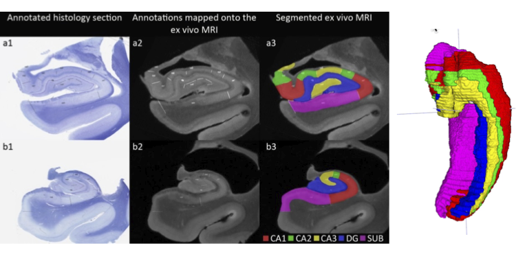
Labeling of hippocampal subfields in an ex vivo MRI scan on the basis of micro-anatomical features derived from histology. Boundaries drawn in histology space are mapped into the MRI space after histology-MRI registration, and volumetric labeling of hippocampal subfields is performed in MRI space.
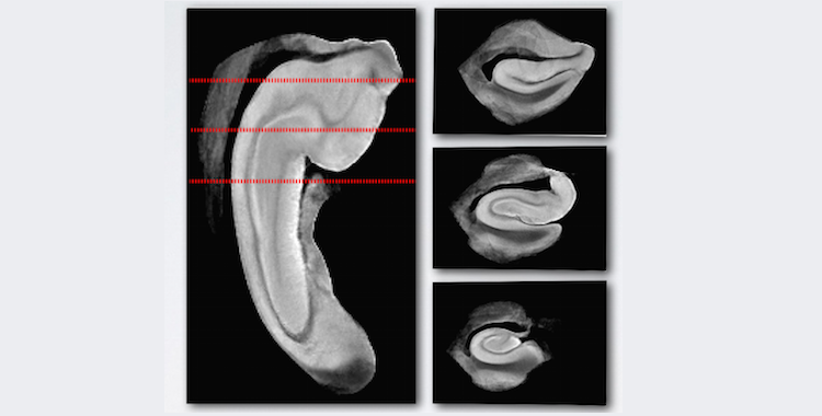
A template generated by volumetric registration of 26 ex vivo MRI scans guided by surface correspondences.